



Metabolic Characteristics and Metabolite Identification (MetID) Strategies for PROTAC Drugs
-

Articles
-

Oct 10, 2026
Introduction to PROTACs
Proteolysis-targeting chimeras (PROTACs) are an emerging class of therapeutic agents that have attracted significant attention from academia, industry, and investment in the field of biomedicine. By expanding the coverage of druggable targets, PROTAC drugs have the potential to solve the problem of drug resistance associated with traditional small molecule inhibitors in the field of targeted protein degradation. In October 2021, the Health and Environmental Sciences Institute (HESI) brought together more than 150 pharmaceutical scientists, regulators, and academic researchers to deeply explore important issues such as the safety evaluation of targeted protein degraders[1]. Participants generally agreed that the safety challenges of PROTACs are mainly related to off-target protein degradation, differences in pharmacokinetics/pharmacodynamics (PK/PD) from conventional small molecule drugs, and the selection of appropriate species for safety assessment in non-clinical trials.
Therefore, there is an urgent need to study the metabolites of PROTACs in depth as early as possible, which is essential for analyzing the distribution of drugs in human circulation, assessing their safety, comparing metabolic differences between different species, and selecting appropriate toxicological species. In this article, we initially discuss the importance of metabolite identification (MetID) at various stages of PROTAC drug development. Subsequently, we summarize the structural and metabolic characteristics of PROTACs. Finally, we outline the challenges encountered during PROTAC MetID research and suggest our corresponding strategies based on our extensive experience. We aim to lay a foundation for developing robust MetID research strategies, help medicinal chemists design PROTAC molecules with favorable pharmacokinetic (PK) attributes, and provide effective direction for the development of PROTAC drugs.
Why Is Metabolite Identification (MetID) Critical for PROTAC Drugs?
Given the relatively large molecular weight of PROTACs, enhancing permeability and solubility often necessitates the incorporation of heteroatoms into the structure during drug design. However, this approach will escalate the risk of introducing more metabolic soft spots. Specifically, the PROTAC linker can undergo metabolic reactions, such as dealkylation or hydrolysis, leading to the production of cleavage products[2]. These metabolites have the potential to competitively bind with the target of PROTACs, thereby inhibiting the pharmacodynamic effects of these drugs, or leading to off-target toxicities. For example, E3 ligase ligand fragments obtained by linker cleavage can bind to non-target proteins as molecular glues and produce off-target effects[1]. Results obtained from in vitro metabolite identification studies can help identify the metabolic soft spots of PROTACs and screen for linker-cleaved products, which further guides the structural optimization of PROTACs to obtain more metabolically stable compounds. In addition, because the metabolic characteristics of a PROTAC cannot be predicted from one of the ligands used for its design and synthesis, PROTACs represent independent chemical entities[2].
During the preclinical development phase, conducting in vitro metabolite identification studies in different species can help assess the metabolic differences of PROTACs between species and select species suitable for toxicological studies. At the same time, the in vitro and in vivo metabolic correlation of PROTACs can be assessed by comparative analysis with the results of in vivo MetID studies, and then PROTAC metabolism in humans can be predicted. Additionally, by analyzing the concentration of a PROTAC and its metabolites in plasma and tissues such as the liver, kidney, and tumor, we can evaluate the in vivo efficacy and toxicity of the PROTAC. This is crucial to understand the potential off-target risks associated with PROTACs[3]. In the Phase I clinical stage, MetID studies can help to analyze the metabolic pathways and clearance of PROTACs in humans and can be used to assess whether the selection of toxicological species for preclinical studies is appropriate (Figure 1).
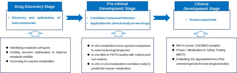
Figure 1. The Role of Metabolite Identification Studies in Various Stages of PROTAC Drug Development
Hence, it is crucial to undertake metabolite identification studies of PROTAC as early as feasible to aid in drug discovery and safety evaluations. The experimental methods and analytical techniques required for PROTAC MetID are more demanding than those for traditional small-molecule drugs. While PROTAC falls under the category of small molecule compounds, its metabolic characteristics significantly diverge from traditional small molecule drugs, attributable to its larger molecular weight and complex structural features.
Structural Characteristics of PROTAC Drugs
PROTAC drugs bring significant therapeutic advantages with their unique structure and mechanism of action. However, this particular structure also endows them with unique physicochemical properties as well as metabolic characteristics that differ from those of conventional small molecule drugs. For a deeper understanding of their metabolic profiles, firstly, we will introduce the structural characteristics of PROTACs, providing a base to summarize and investigate their metabolic characteristics. A PROTAC primarily consists of three critical structures: one region is the ligand structure that targets the protein of interest (POI); another region is the ligand structure for an E3 ligase; and situated between these two is the linker, which connects both ligands (Figure 2). In the following sections, we will individually introduce these three components based on their structural characteristics.
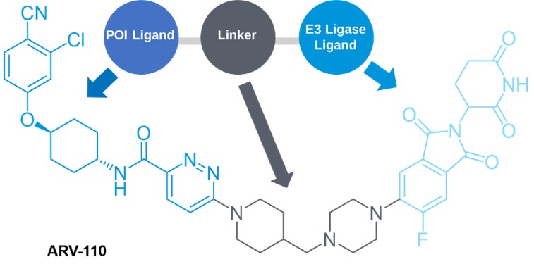
Figure 2. Structural Diagram of PROTAC and Representative Structure of PROTAC: ARV-110
POI Ligand
Among the reported PROTAC molecules to date, the number of POI ligands exceeds 360[4]. The target proteins include androgen receptor (AR), estrogen receptor (ER), bromodomain-containing protein (BRD), and Bruton tyrosine kinase (BTK). The POI ligand component of PROTAC typically utilizes active inhibitors that are either marketed or reported in scholarly literature[3]. For instance, Gray's group, in 2019, developed BSJ-03-204[5], derived from the CDK4/6 inhibitor Palbociclib. In 2021, Bian's team developed B03[6], which is based on the CDK9 inhibitor BAY-1143572.
PROTAC drugs target not only conventional small molecule drug targets but also extend to non-druggable proteins, scaffold function proteins, protein polymers, drug-resistant mutant proteins, and specific isoform proteins. For example, signal transducer and activator of transcription 3 (STAT3) is a transcription factor closely related to the development of cancer and is considered to be a non-druggable protein. Based on SI-109, a binder of the SH2 domain in STAT3, Wang's group[7] developed the PROTAC molecule SD-36 which has high degradation activity for the STAT3 protein in both in vitro and in vivo experiments (Figure 3). KT-333, also a PROTAC drug targeting STAT3, developed by Kymera, has entered phase I clinical trials to assess its safety in patients with large granular lymphocytic leukemia.
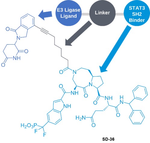
Figure 3. STAT3 Degradant SD-36
E3 Ligand
There are over 600 known types of E3 ligases encoded by the human genome. However, in PROTAC-related drug design, the primary E3 ubiquitin ligases include cereblon (CRBN, with 71 subtypes), von Hippel-Lindau (VHL, with 47 subtypes), cellular inhibitor of apoptosis protein (cIAP, with 7 subtypes), and murine double minute2 (MDM2, with 4 subtypes) (Figure 4)[3]. Among PROTACs that have entered the clinical stage and have published structures, more than 39% of the molecules have CRBN ligands, and only DT2216 contains a VHL ligand.
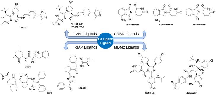
Figure 4. Representative E3 Ligands
Investigating novel E3 ubiquitin ligase targets is a prominent area of interest in PROTAC research. In recent years, several structurally unique E3 ligases have been discovered, including DCAF11, DCAF15, DCAF16, RNF114, RNF4, AhR, FEM1B, and KEAP1. Additionally, over 80 novel E3 ligase ligands have been reported. Moving forward, it is anticipated that researchers will identify a more diverse range of E3 ligases, as well as E3 ligase ligands with smaller molecular weight and higher activity.
Linker
The PROTAC linker, an important part, connects the two active groups of PROTAC drugs, playing an essential role in the activity, metabolic stability, and kinetic properties of PROTAC molecules. Over 1500 different linker structures have been reported[4], and from a molecular variability perspective, linkers can be divided into two types: flexible and rigid (Figure 5). Relevant literature[2] indicates that neither excessively short nor excessively long linkers are beneficial for the efficacy of a PROTAC. If the linker is too short, it can impede the binding of ligands on both sides to the corresponding protein due to steric hindrance, making the formation of the ternary complex difficult. Conversely, when the linker is too long, the E3 ligase cannot sufficiently approach the target protein, preventing the ubiquitination process from occurring, and the excessive molecular weight reduces the PROTAC's membrane permeability. Studies suggest that linkers possessing rigid elements (e.g., a piperidine moiety) or polar groups (e.g., polyethylene glycol) can enhance the pharmacokinetic properties of PROTACs.
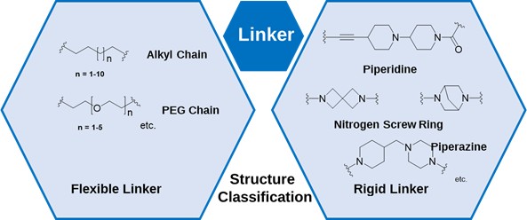
Figure 5. Representative Structures of Linkers Used in PROTACs
Besides the structure of the linker, its attachment site can also influence the selectivity and activity of PROTAC. Generally, the principles for selecting ligation sites are as follows: 1) Do not diminish the binding capacity between E3 ligase ligands or POI ligands and their receptors; 2) Select the solvent-exposed region of the ligand binding pocket. 3) Ensure the integrity of POI ligands as much as possible when attaching the linker to prevent any impact on their binding capacity. Figure 6 illustrates the common ligation sites of E3 ligase ligands (indicated by the red arrow section) as well as the ligation sites of several POI ligands[3].
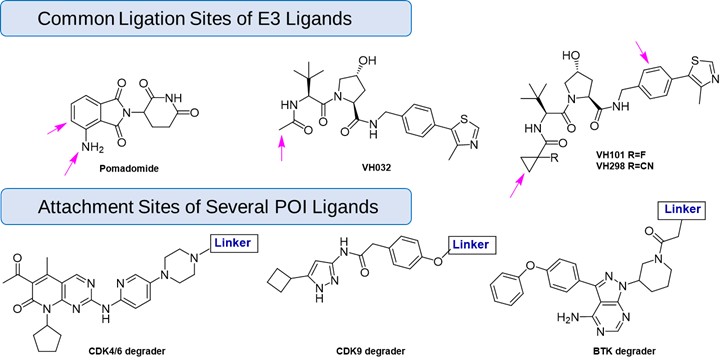
Figure 6. Common Attachment Sites of Linkers with E3 and POI Ligands
Metabolic Characteristics of PROTAC Drugs
Due to their unique structural characteristics, the molecular weight and physicochemical properties of PROTAC often exceed the limitations of Lipinski's Rule of Five, and its metabolic process is also significantly different from that of traditional small molecule drugs. An in-depth understanding of their metabolic properties will help researchers to more effectively cope with the challenges encountered in the study of metabolite profiling. Therefore, this part will thoroughly summarize the metabolic characteristics of PROTAC drugs based on their structural features.
The Influence of the E3 Ligand on PROTAC Metabolism
Cruciani's team[2] discovered, through comparing three data sets, that for PROTACs targeting BET, CK2, and PARP proteins, when the POI ligands remain constant and linkers are similar, the metabolic stability of PROTACs with thalidomide as the E3 ligase ligand is significantly lower than those with VHL ligand (as depicted in Figure 7). The team hypothesizes that the plausible reason could be that thalidomide and its analogs contain multiple amide structures, which might undergo non-enzyme catalyzed hydrolysis reactions. Another possibility is that due to the larger molecular weight of the VHL ligand, the corresponding PROTAC has reduced permeability[3], thereby not providing an accurate estimation of its half-life. Therefore, for metabolic stability assays of PROTACs containing VHL ligands, liver microsomes may be a more appropriate in vitro metabolic study system[8].
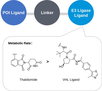
Figure 7. Effect of E3 Ligands on the Metabolic Stability of PROTACs
The Influence of the POI Ligand on PROTAC Metabolism
Cruciani's team[2] discovered that the metabolic stability of PROTAC molecules changes as the POI ligand varies, keeping the linker and E3 ligase ligand constant. Figure 8 illustrates that for PROTACs using the AR antagonist as the POI ligand, their metabolic stability is lower compared to other POI ligands, regardless of changes in the linker and E3 ligase ligand. Metabolite identification studies identified that the primary soft metabolic spots of these compounds are on the POI ligand, not on the E3 ligase ligand. Given the fast metabolic rate of the POI ligand itself (half-life of 18.3 min), this suggests that the metabolic stability of the POI ligand needs to be considered when designing PROTAC drugs to avoid significant weaknesses. For example, the PROTAC drug ARV-110, uses an AR antagonist with better metabolic stability than the POI ligand, boasting a half-life of up to 110 hours.
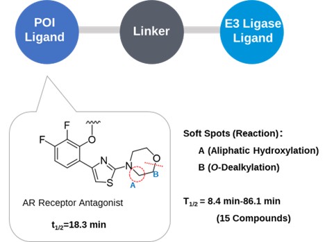
Figure 8. Effect of POI Ligands on Metabolic Stability
The Influence of the PROTAC Linker on Metabolism
In PROTAC molecules, the linker is the section most prone to be metabolized. The main metabolic sites are located where the linker attaches to the ligand. Variations in the linker's length, attachment points, and degree of rigidity or flexibility can significantly influence the overall metabolic stability of the molecule.
Effect of Linker's Length and Ligation Site on the Metabolic Stability of PROTAC
For the majority of PROTACs, the metabolic stability of the molecule tends to diminish as the linker length extends. As depicted in Figure 9, R1 represents a PROTAC, ingeniously designed using JQ1 (a BET inhibitor) and Thalidomide. When the straight-chain alkyl linker in R1 is elongated from four methylene units to eight, forming R2, the half-life dramatically contracts from 135 minutes to just 18.2 minutes. This phenomenon can likely be attributed to the shorter linker's increased steric hindrance, which effectively prevents the PROTAC from accessing the metabolic enzyme's catalytic site.
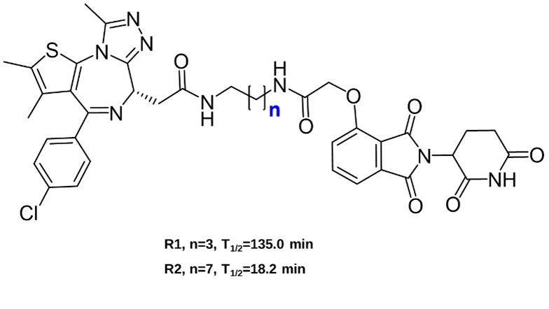
Figure 9. Effect of Linker Length on the Metabolic Stability of PROTACs
It has also been found that the changes in the ligation site of linker and E3 ligase ligand, may also affect the metabolic stability of PROTAC, as shown in Figure 10.
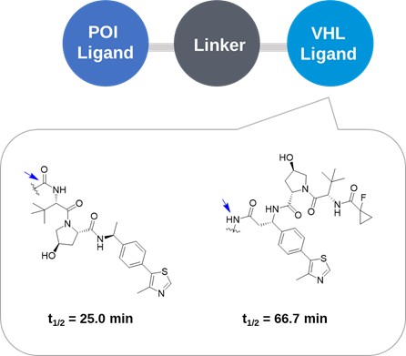
Figure 10. Impact of the Linker Attachment Site on the Metabolic Stability of PROTACs
Effect of Linker Rigid Flexibility on the Metabolic Stability of PROTAC
Studies have shown that when both the POI (Protein of Interest) ligand and the E3 ligase ligand are kept constant, the assembly of the PROTAC molecule with different linkers can significantly impact its metabolic stability (as depicted in Figure 11). Specifically, PROTACs featuring piperidine or triazole ring linkers demonstrate a prolonged half-life compared to those with straightforward simple linear linkers. This evidence suggests that PROTACs constructed with rigid linkers tend to exhibit enhanced metabolic stability.
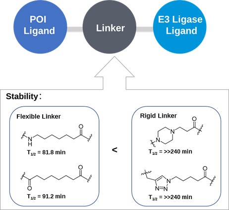
Figure 11. Effect of Linker Rigidity on Metabolic Stability
Incorporating rigid structures into the linker presents a superior approach to bolster metabolic stability. For instance, in the development process of ARV-110 (as depicted in Figure 12), the initial flexible chain-like linker was refined into a stiff cyclic linker, bridged by piperidine and piperazine rings. This optimization notably enhanced its metabolic stability and therapeutic efficacy. Arvinas, the company that developed ARV-110, implemented the same linker design in another PROTAC drug, ARV-471[2].
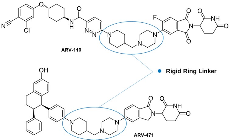
Figure 12. Structure of ARV-110 and ARV-471 with Rigid Cyclic Linker
In 2021, Wang Yonghui's research team at Fudan University[9] unveiled the PROTAC molecule 6e. Despite its high degradation activity against BTK protein, 6e demonstrated relatively low metabolic stability, with a half-life of just 1.3 minutes in mouse liver microsomes. However, in 2023, the team significantly enhanced the molecule's metabolic stability by implementing a strategy to increase the rigidity of its linker. Through conducting structure-activity relationship studies on a series of rigid linkers and CRBN ligands, they replaced the linear polyethylene glycol linker with a more rigid one, featuring two piperidine rings. This modification led to the creation of molecule 3e, which not only displayed a greatly improved metabolic stability, with a half-life exceeding 145 minutes in mouse liver microsomes but also outperformed its predecessor, 6e, in terms of activity (as depicted in Figure 13).
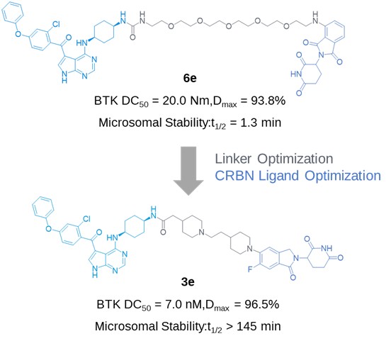
Figure 13. Study Case: Enhancing PROTAC Metabolic Stability and Activity through Structural Optimization
Relationship Between the Metabolic Characteristics of Various PROTAC Components and the Whole Molecule
Considering that a PROTAC molecule is constituted by three crucial structures, one might wonder if it's possible to predict the metabolic characteristics of PROTAC based on the metabolic traits of each ligand. To address this, Cruciani's team[2] embarked on a comprehensive study. As demonstrated in Figure 14, the POI ligand and CRBN ligand individually undergo metabolic reactions of alkyl hydroxylation and amide hydrolysis, respectively. However, when these ligands are conjugated via a linker into a PROTAC molecule, neither ligand exhibits the respective metabolism, with the primary metabolic activity occurring on the linker itself. Moreover, the metabolic rate of the entire PROTAC molecule varies from that of its separate ligands. Thus, as a novel chemical entity, PROTAC's metabolic characteristics cannot be extrapolated merely from the metabolic attributes of its constituent parts, underscoring the need for a holistic investigation of its metabolites through robust MetID.
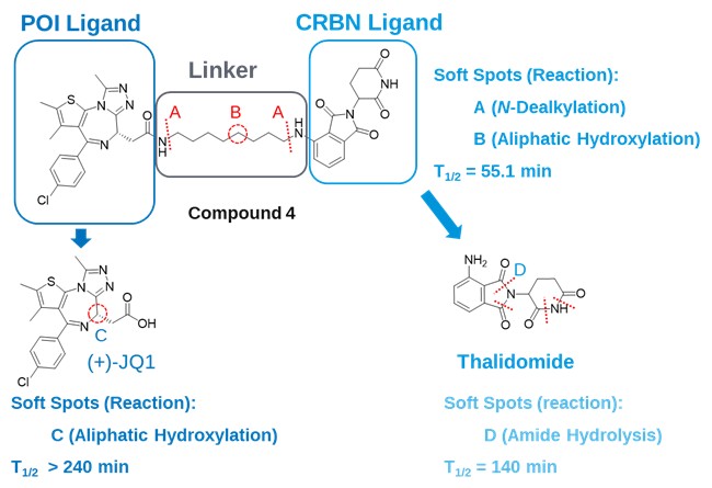
Figure 14. Comparison of PROTAC's Metabolic Soft Spots with Those of Its Individual Ligands
Summary of Metabolic Characteristics of PROTACs
From the above analysis, it can be seen that the type of E3 ligase, the stability of the POI ligand, and the length, ligation site, and rigidity of the linker all affect the metabolism of PROTAC. The metabolic characteristics of PROTAC molecules are different from the addition of metabolic characteristics of their various partitions, and the metabolic rate may also be completely different.
Metabolic pathways of PROTACs include hydroxylation of its ligand moiety, amide hydrolysis, O-dealkylation, and hydroxylation of the linker moiety, the hydroxylation, N-dealkylation, and amide hydrolysis of the linker moiety. If the drug contains a PEG-like linker, it is highly susceptible to O-dealkylation reactions. Enzymes involved in the metabolism of PROTACs mainly include phase I metabolizing enzymes (CYP enzymes) and common phase II metabolic enzymes (uridine diphosphate glucuronosyltransferases (UGTs) and sulfotransferases (SULTs), etc.). In addition, Cruciani's team[2] found that for PROTACs containing VHL ligands, aldehyde oxidase (hAOX) is involved in metabolism and catalyzes the hydroxylation of its thiazole ring. Since aldehyde oxidase is more highly expressed in humans, metabolic studies targeting this enzyme require special attention.
What Are the Challenges and Strategies for PROTACs in Metabolite Profiling and MetID?
The process of identifying metabolites for a PROTAC molecule aligns closely with that associated with traditional small-molecule drugs (Figure 15). Initially, an appropriate metabolic study system is selected for either in vitro incubation or in vivo administration in animals. Following this, samples, either from incubation or collected in vivo, undergo a process of LC-UV-HRMS analysis after protein precipitation, centrifugation, supernatant transfer, nitrogen blowing, concentration, and redissolution. The parent drug and its metabolites are separated through chromatography, and a comparison of the information between the drug substance and its metabolites is conducted based on the MS1 and MS/MS signals from high-resolution mass spectrometry, the metabolic pathways, and reaction sites of PROTACs are analyzed.
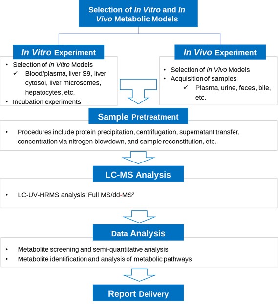
Figure 15. Workflow for Metabolite Identification of PROTACs
However, due to the distinct structural and metabolic characteristics of PROTAC drugs compared to traditional small molecules, the study of their metabolites presents additional challenges. These include the selection of appropriate in vitro and in vivo models, addressing issues of non-specific adsorption and solubility of parent drugs during sample preparation, and efficiently acquiring the relative abundance of metabolites. Furthermore, accurately completing the structural characterization in the data analysis phase also poses a challenge.
Drawing from the extensive research expertise of the WuXi AppTec DMPK (Drug Metabolism and Pharmacokinetics) MetID Team, we have devised a comprehensive research strategy to effectively address these challenges encountered during PROTAC metabolite identification studies (Figure 16). Our approach aims to deliver high-quality research reports to our clients, thus expediting the drug development process for PROTAC molecules. We delve into these strategies in greater detail in the following sections.
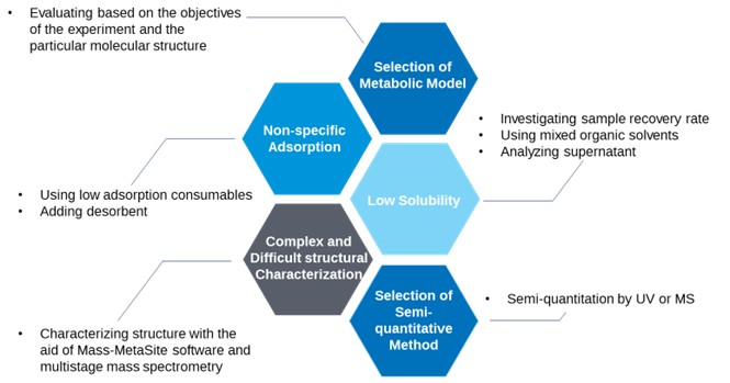
Figure 16. Diagram of Challenges and Strategic Solutions for PROTAC Metabolite Identification Studies
Selection of Metabolic Models for In Vitro Metabolite Identification
The selection of an appropriate metabolic model is key for successful in vitro and in vivo metabolite research. For the study of PROTAC molecules, from which dimensions should appropriate metabolic models be selected?
The selection of an appropriate in vitro metabolic system for studying PROTAC molecules depends on the experiment’s objective and the molecule’s structural characteristics (Table 1). If the study involves the metabolic stability of PROTAC under the action of blood hydrolytic enzymes, in vitro plasma or blood incubation can be utilized. Liver S9 or liver cytosol is selected as the metabolic system when aldehyde oxidase is involved in metabolism. This is particularly critical for PROTACs containing a VHL ligand, as its thiazole ring may be hydroxylated under aldehyde oxidase catalysis[2]. Hepatocyte systems are used to study the effects of CYP enzymes and non-Phase I metabolizing enzymes on compounds. For larger molecular weight PROTAC molecules with poor cell permeability, the liver microsomal system is more suitable than the hepatocyte system for studying reactions involving only CYP enzymes during a metabolic stability assay[8].
For in vitro-in vivo metabolism correlation, plasma, urine, feces, bile, and other in vivo samples can be used to evaluate the correlation between in vitro and in vivo metabolism of PROTAC, helping predict human metabolism. Metabolite identification studies of human in vivo samples can assess the intrabody systemic exposure scenarios of a PROTAC and determine the appropriateness of the selected toxicological species.
Table 1. The in vitro and in vivo systems for PROTAC metabolism and their respective purposes
Study Subjects | Matrix | Purpose |
PROTAC | In vitro blood/plasma | Investigating hydrolases-mediated metabolism in blood/plasma |
Liver S9/Liver cytosol (With or without aldehyde oxidase inhibitor) | Investigating aldehyde oxidase-mediated metabolism | |
Liver microsomes | Investigating CYP enzyme-mediated metabolism | |
Hepatocytes | Investigating both phase I and II enzyme-mediated metabolism | |
In vivo samples (Plasma, blood, urine, feces, bile) | Investigating the correlation between in vitro and in vivo metabolism to predict metabolic processes in the human body | |
In vivo samples obtained from the human body | Evaluating the exposure of PROTACs in the human body and the appropriateness of species selection for toxicology studies |
Method Development for Metabolite Profiling
During the method development of PROTAC molecules, non-specific adsorption, and solubility issues are common challenges. Improper handling of these issues can significantly impact the comprehensiveness and accuracy of metabolite analysis results. Hence, it is crucial to develop methods and strategies in advance to effectively address these challenges.
For non-specific adsorption issues, the large molecular weight of PROTAC can result in easy non-specific binding to the surface of consumables[10]. To mitigate this, it's recommended to use low-adsorption consumables and assess compound adsorption during the method development stage. If a compound is found to bind non-specifically to consumables, the addition of an appropriate amount of desorbent may be required.
To address solubility issues, the solubility can be first evaluated by investigating the recovery of PROTAC and selecting the suitable solvent. If the recovery rate is insufficient, it may result in missed metabolites, inaccurate semi-quantitative results, and ultimately unreliable metabolite identification. For instance, a PROTAC molecule in a mixed acetonitrile/water solvent may show low recovery at 0 min of hepatocyte incubation (T0 sample) and cause system stratification. Adjusting the proportion of acetonitrile might not improve this situation, suggesting the need for other organic reagents in the reconstitution solution. In one case, the highest recovery rate was achieved when the reconstitution solution was a mixed solvent made up of several organic reagents and water, fulfilling the analytical requirements (as depicted in Figure 17).
To solve PROTAC's solubility problem, strategies may include using solutions containing mixed organic reagents for compound dissolution, dilution, and pretreatment, or directly using the supernatant after protein precipitation and centrifugation for LC-UV-HRMS analysis. By adopting these strategies, the problems of non-specific adsorption and solubility of PROTAC samples can be effectively overcome to ensure the comprehensiveness and accuracy of metabolite profiling and identification results.
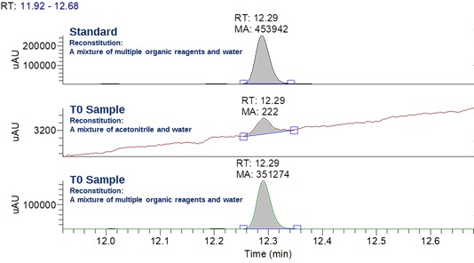
Figure 17. LC-UV Chromatograms of a PROTAC in Different Reconstitution Solvents
Structural Characterization of Metabolites
Structural characterization is a critical step in metabolite identification. For PROTAC molecules, their large molecular weight and the presence of multiple reaction sites pose significant challenges for efficient and high-quality structural characterization, a crucial aspect of metabolite research. To solve these challenges, we've developed a set of strategies specifically for PROTAC metabolite profiling and structure identification (Figure 18). These strategies, derived from extensive experimental explorations and experiences, leverage the strengths of the metabolite identification software Mass-MetaSite, the expertise of senior scientists, and high-resolution mass spectrometry platforms.
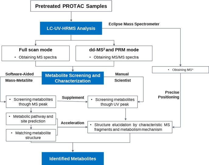
Figure 18. Workflow for the Metabolite Identification of PROTACs
In this strategy, our experienced professionals characterize metabolite structures by interpreting the UV and mass spectral information from LC-UV-HRMS analysis data. This includes elucidating the metabolic pathways and metabolic sites of PROTAC metabolites through chromatographic peak searching and mass spectral fragment information interpretation. Simultaneously, we employ Mass-MetaSite software for automated screening and identification of metabolites, calculating metabolic pathways and potential metabolic sites, and structurally aligning them with experimental results. This integrated approach that combines expert interpretation with automated software analysis speeds up the process of characterizing PROTAC metabolite structures. This assists researchers and developers in their work, making the process more efficient and reliable.
Additionally, WuXi AppTec DMPK platform features an industry-leading Thermo Scientific™ Orbitrap Eclipse™ Tribrid™ mass spectrometer capable of multi-stage (MS3 and above) mass spectrometry data. During the structure characterization process, R&D scientists utilize this multi-stage mass spectrometry data to deeply analyze the fragmentation behavior of drug candidates and their metabolites. This allows for the acquisition of more refined fragment information and precise localization, ensuring high-quality structural characterization.
Through our strategy that combines professional software, experienced expert teams, and industry-leading high-resolution mass spectrometry platforms, structural characterization in MetID can be efficiently carried out. This approach helps us navigate the challenges posed by the complexity of PROTAC molecules, speeds up PROTAC metabolite research, and provides crucial support for the design and development of PROTAC drugs.
Semi-quantification of Metabolites
Through semi-quantitative analysis of characterized metabolites, we can estimate the relative abundance of the parent drug and its metabolites. This allows for an initial understanding of the proportion of each metabolite and helps identify the main metabolites. This process is crucial for identifying metabolic soft spots, comparing in vitro metabolic differences across species, and comparing in vitro and in vivo metabolic differences. The information generated provides a basis for further structural optimization of subsequent drugs, prediction of drug metabolism in the human body, and safety evaluation of metabolites.
Given the structural properties of PROTACs, it is vital to ensure the accuracy of semi-quantitative results[11]. In semi-quantification using MS, the difference in ionization efficiency between the parent drug and its metabolites in the ion source can impact the semi-quantitative data accuracy. Moreover, PROTACs and their metabolites often produce not only singly charged ions, but also multiply charged ions and in-source fragments. These factors need to be fully considered when performing semi-quantification by mass spectrometry integration. This involves synchronously extracting and integrating singly charged ions, multiply charged ions, and in-source fragments, which enhances the accuracy of semi-quantification. For instance, in a PROTAC study, we integrated the extracted ion current for a metabolite. The peak area after superimposing the extracted singly and multiply charged ions was approximately eight times that of the singly charged extracted ion.
When employing a UV detector for semi-quantification, it's crucial to note that if the E3 ligand of a PROTAC has a different UV chromophore compared to the POI ligand, the metabolite resulting from linker breakage might differ from the parent drug in terms of absorbance coefficient. This discrepancy could significantly reduce the accuracy of UV semi-quantitative data. In such scenarios, other detectors, such as CAD (Charged Aerosol Detector), can be considered for more accurate results.
Therefore, the approach to semi-quantifying metabolites should be chosen comprehensively, taking into account their structural characteristics, UV spectra, and mass spectra.
Summary
After over two decades of development, PROTACs have gained favor among leading pharmaceutical companies and research institutions worldwide due to their unique mechanism of action and advantages. Metabolite studies are crucial during the discovery, preclinical, and clinical stages of PROTAC drugs, making the experimental data for metabolite identification increasingly critical for new drug applications. Leveraging the advanced Waters ACQUITY UPLC liquid phase system and mass spectrometry systems such as Orbitrap EclipseTM TribridTM and Q-ExactiveTM; HF, in conjunction with data processing software like Mass-MetaSite, Compound Discoverer, and MetaboLynx, WuXi AppTec DMPK metabolite identification team has developed a research platform for PROTAC metabolite profiling and identification. This platform, based on Ultra Performance Liquid Chromatography-High Resolution Mass Spectrometry (UPLC-UV-HRMS) technology, has established a series of robust solutions to overcome the challenges in the experimental process, thereby aiding the metabolic study of PROTAC molecules for numerous drug development companies globally. Looking forward, we anticipate an increasing number of PROTAC drugs transitioning into clinical studies and, ultimately, achieving a successful market launch, bringing hope to more patients.
WuXi AppTec DMPK offers integrated pharmacokinetics services spanning early discovery, preclinical development, and clinical stages, supported by research centers in China (Shanghai, Suzhou, Nanjing, and Nantong) and the United States (New Jersey) to help accelerate your drug development. With a team of over 1,000 scientists serving more than 1,700 customers worldwide and over fifteen years of regulatory filing experience, WuXi AppTec DMPK has successfully supported more than 2,100 Investigational New Drug (IND) applications.
Authors: Hong Zhang, Ruixing Li, Weiqun Cao
Talk to a WuXi AppTec expert today to get the support you need to achieve your drug development goals.
Committed to accelerating drug discovery and development, we offer a full range of discovery screening, preclinical development, clinical drug metabolism, and pharmacokinetic (DMPK) platforms and services. With research facilities in the United States (New Jersey) and China (Shanghai, Suzhou, Nanjing, and Nantong), 1,300+ scientists, and over fifteen years of experience in Investigational New Drug (IND) application, our DMPK team at WuXi AppTec are serving 1,700+ global clients, and have successfully supported 2,100+ IND applications.
Reference
[1] Jones, L. H.; Mitchell, C. A.; Loberg, L. et al. (2022). Targeted protein degraders: a call for collective action to advance safety assessment. Nat Rev Drug Discov. 21 (6): 401-402.
[2] Goracci, L.; Desantis, J.; Valeri, A. et al. (2020). Understanding the Metabolism of Proteolysis Targeting Chimeras (PROTACs): The Next Step toward Pharmaceutical Applications. J Med Chem. 63 (20): 11615-11638.
[3] Cao, C.; He, M.; Wang, L. et al. (2022). Chemistries of bifunctional PROTAC degraders. Chem Soc Rev. 51 (16): 7066-7114.
[4] Weng, G.; Cai, X.; Cao, D. et al. (2023). PROTAC-DB 2.0: an updated database of PROTACs. Nucleic Acids Res. 51 (D1): D1367-D1372.
[5] Jiang, B.; Wang, E. S.; Donovan, K. A. et al. (2019). Development of Dual and Selective Degraders of Cyclin-Dependent Kinases 4 and 6. Angew Chem Int Ed Engl. 58 (19): 6321-6326.
[6] Qiu, X.; Li, Y.; Yu, B. et al. (2021). Discovery of selective CDK9 degraders with enhancing antiproliferative activity through PROTAC conversion. Eur J Med Chem. 211: 113091.
[7] Bai, L.; Zhou, H.; Xu, R. et al. (2019). A Potent and Selective Small-Molecule Degrader of STAT3 Achieves Complete Tumor Regression In vivo. Cancer Cell. 36 (5): 498-511.
[8] Khojasteh, S. C.; Argikar, U. A.; Driscoll, J. P. et al. (2021). Novel advances in biotransformation and bioactivation research - 2020 year in review. Drug Metab Rev. 53 (3): 384-433.
[9] Chen, S.; Chen, Z.; Lu, L. et al. (2023). Discovery of novel BTK PROTACs with improved metabolic stability via linker rigidification strategy. Eur J Med Chem. 255: 115403.
[10] Fandozzi, C.; Evans, C.; Wilson, A. et al. (2019). 2019 White Paper on Recent Issues in Bioanalysis: Chromatographic Assays (Part 1 - Innovation in Small Molecules and Oligonucleotides & Mass Spectrometric Method Development Strategies for Large Molecule Bioanalysis). Bioanalysis. 11 (22): 2029-2048.
[11] Pike, A.; Williamson, B.; Harlfinger, S. et al. (2020). Optimising proteolysis-targeting chimeras (PROTACs) for oral drug delivery: a drug metabolism and pharmacokinetics perspective. Drug Discov Today. 25 (10): 1793-1800.
[12] Liang Shen, editor. Drug Metabolism and Pharmacokinetics: Frontiers, Strategies, and Applications. Wiley. 2025. http://doi.org/10.1002/9781394300150.


Related Services and Platforms


-
 MetID (Metabolite Profiling and Identification)Learn More
-
Novel Drug Modalities DMPK Enabling PlatformsLearn More
-
In Vitro MetID (Metabolite Profiling and Identification)Learn More
-
In Vivo MetID (Metabolite Profiling and Identification)Learn More
-
Metabolite Biosynthesis and Structural CharacterizationLearn More
-
Metabolites in Safety Testing (MIST)Learn More
-
PROTAC DMPK ServicesLearn More
-
ADC DMPK ServicesLearn More
-
Antibody-Oligonucleotide Conjugate (AOC) DMPK ServicesLearn More
-
Oligo DMPK ServicesLearn More
-
PDC DMPK ServicesLearn More
-
Peptide DMPK ServicesLearn More
-
mRNA DMPK ServicesLearn More
-
Covalent Drugs DMPK ServicesLearn More



Stay Connected
Keep up with the latest news and insights.

