

Quantitative Whole-body Autoradiography (QWBA)
After the animals are administered with radiolabeled compound, quantitative whole-body autoradiography (QWBA) is applied to obtain the distribution of total radioactivity in the whole body, and complete and detailed tissue distribution results are obtained to provide a theoretical basis for pharmacodynamic and toxicological studies.

-
Overview
-
Service
-
Study Strategies
-
Case Study
-
Instruments
-
FAQs
-
Related Resources
-
Related Services


Overview
The obvious advantage of the QWBA studies is to assess whether all the drug related materials can be completely eliminated from the body, without the information of metabolites derived from the drug candidates. QWBA method can be used in a variety of animal models, including mice, rats, NHPs, etc. QWBA image also provides valuable drug distribution information for small tissues, such as brain, eyes, even for fetus and tumors, etc. Another important application of QWBA is to assess melanin associated binding in pigmented rodents (like pigmented Long-Evans rats), and calculate human dosimetry dosage from the tissue distribution data.
Learn More


Service


Study Strategies
QWBA is a technique for imaging the distribution and time course of elimination of a radiolabeled compound and its metabolites, commonly performed in rodents (rat is mostly used). Because it generally takes weeks for sample processing and measurement, long-lived radiolabels such as 14C and 3H are often chosen for the study. A typical experimental design for QWBA study is as below: each rat receives a single dose of a radiolabeled drug, 5-8 time points, often only male, individual animals at each time point, and the collection time is longer than traditional tissue distribution, in order to obtain a complete tissue elimination rule.
Case Study
-
-

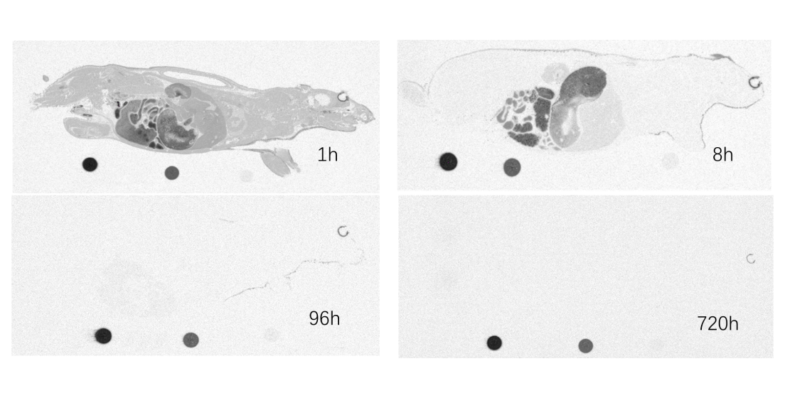

Representative whole-body autoradiography showing melanin in the uveal tract of [14C] compound
Melanin is a pigment found in skin, the uveal tract, and hair. Melanin binding of radiolabeled compound causes long exposure of radiation to pigment tissues and further damages tissue especially in eye. One of the important applications of QWBA is to measure melanin-associated binding in pigmented rats and to predict potential adverse effects to human volunteers participating in a human radiolabeled AME study. The pictures as below showed that [14C] compound -derived radioactivity was obviously associating with melanin in the uveal tract, radioactivity level was still high at 720h post-dose.
-
Instruments
-
-

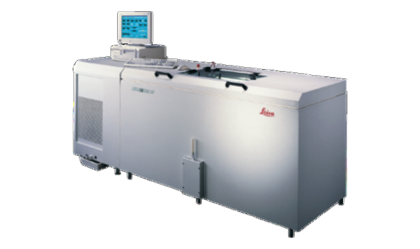
Cryostat Microtome
-
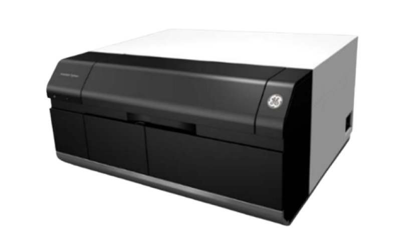
Multifunctional Laser Imager
-
FAQs
-

What are the key benefits of QWBA?


First, to track how compounds of interest are distributed in the body, even for small tissues; second, to assess melanin associated binding in pigmented rodents (like pigmented Long-Evans rats), and calculate human dosimetry dosage from the tissue distribution data.
-
What are the application scenarios of QWBA technology?
In addition to the general tissue distribution studies, QWBA technology also can be used in placental transfer study, tumor distribution study, etc.
-
What animal models can QWBA be used in?
QWBA method can be used in a variety of animal models, including mice, rats, NHPs, etc.
Related Resources


-

List of Biological Sample Collection of Quantitative Whole-body Autoradiography (QWBA)
 BrochuresAug 21, 2024Learn More
-
Quantitative Whole-body Autoradiography (QWBA) Enables ADC Tissue Distribution Studies in Tumor-bearing Nude Rodents
BlogsAug 18, 2023Learn More -
Quantitative Whole-body Autoradiography (QWBA) vs. Mass Balance Studies
BlogsMay 04, 2023Learn More -
Tissue Distribution Using Quantitative Whole Body Autoradiography (QWBA) of [14c]-Paclitaxel in Tumor Bearing Mice
PostersMay 04, 2023Learn More


Stay Connected
Keep up with the latest news and insights.
