



PEGylated Peptides: Precursor vs. Product Ion Scan in Peptide Bioanalysis
-

Articles
-

Aug 27, 2026
Why Is PEGylation Important for Peptide Pharmacokinetics?
In the field of biopharmaceuticals, peptide drugs have become one of the most promising areas of global drug discovery owing to their core advantages of high efficacy, high specificity, and high safety. Among the 370 new drugs approved by the FDA from 2016 to 2023, 31 were peptide drugs, second only to monoclonal antibodies. These drugs span multiple therapeutic areas, including diabetes, oncology, and cardiovascular diseases [1]. In particular, driven by GLP-1 drugs, peptide drugs have achieved rapid growth in the metabolic disease field, with a compound annual growth rate of 8.1% from 2018 to 2023. The market size is expected to exceed USD 230 billion by 2030 [2].
However, structural limitations of peptide drugs have continued to constrain their development. Poor stability and weak membrane permeability lead to extremely low oral bioavailability, while short half-lives increase dosing frequency and the risk of adverse reactions. PEG conjugation, also known as PEGylation, provides an effective solution. By covalently attaching PEG chains, this strategy can significantly prolong drug half-life and reduce immunogenicity. For example, Empaveli, approved in 2021, showed a markedly extended half-life after modification, enabling longer dosing intervals [3]. Nevertheless, PEG modification increases the molecular weight of drug molecules from several thousand daltons to tens of thousands of daltons, fundamentally changing the analytical approaches used for conventional peptides. Among these analytical considerations, the choice of mass spectrometric scan mode is particularly critical. This article focuses on two modes: product ion scanning and precursor ion scanning, and discusses efficient analytical strategies for PEGylated peptide drugs.
Clinical Value and Peptide Bioanalysis Challenges of PEGylated Drugs
What Are the Clinical Breakthroughs of PEGylation Technology
PEGylation was first proposed by Abuchowski in 1977. Its core principle is to covalently link polyethylene glycol (PEG) to biomolecules such as peptides and proteins. Through steric hindrance and strong hydration, PEGylation comprehensively optimizes the physicochemical properties and pharmacokinetic behavior of drugs [4]. As of 2024, the FDA has approved four PEGylated peptide drugs, and their significant half-life extension fully validates the clinical value of PEGylation technology.
Table 1. FDA-approved PEGylated peptide drugs
Trade Name | Company | PEGylated Entity | Approved Year | MW of PEGs | Indications | T1/2 |
SYFOVRE (Pegcetacoplan injection) | Apellis | Cyclic peptide (C3 inhibitor) | 2023 | / | Geographic atrophy | ~2.5 h → ~600 h |
Empaveli (Pegcetacoplan) | Apellis | Cyclic peptide (C3 inhibitor) | 2021 | / | Paroxysmal nocturnal hemoglobinuria | ~2.5 h → ~83 h |
Omontys (Peginesatide ) | Takeda | EPO receptor agonist peptide (EPO-mimetic) | 2012 | 2×20 kDa | Anemia | ~0.167 h → ~ 130 h |
Mircera (Methoxy PEG-epoetin beta) | Roche | Epoetin beta (methoxy PEG-epoetin beta) | 2007 | 30 kDa | Anemia | ~5 h → ~130 h |
Notably, Empaveli and SYFOVRE have exactly the same active ingredient. Due to differences in the route of administration and indications, their half-lives reach 83 hours and 600 hours, respectively. This fully demonstrates the substantial potential of PEGylation technology in optimizing dosing frequency and improving patient adherence [5]. Behind these clinical benefits, however, are new analytical challenges introduced by PEG modification. The following sections analyze these challenges and propose corresponding peptide bioanalysis strategies.
What Are the Core Analytical Challenges for PEGylated Peptides
At present, quantitative analytical methods for PEGylated peptides mainly include enzyme-linked immunoassay, radioisotope labeling, nuclear magnetic resonance, and liquid chromatography-tandem mass spectrometry (LC-MS/MS). A comparison of these methods is presented in Table 2.
Table 2. Performance comparison of different quantitative analytical methods for PEGylated peptides
Method / Evaluation Dimension | Sensitivity | Selectivity | Efficiency | Scope of Application |
Enzyme-Linked Immunosorbent Assay (ELISA) | Moderate | High | High | High-throughput screening / Quantitative analysis |
Radioisotope Labeling | High | High | Moderate | Detection of low-concentration samples |
Nuclear Magnetic Resonance (NMR) | Low to moderate | Low | Low to moderate | Structural elucidation of purified samples |
Liquid Chromatography-Tandem Mass Spectrometry (LC-MS/MS) | Very high | Very high | Moderate to high | Qualitative and quantitative analysis of complex samples |
As shown in the table, LC-MS/MS has become the preferred analytical method for DMPK studies due to its core advantages of high sensitivity, high selectivity, and compatibility with complex samples. However, four key challenges remain in the analysis of PEGylated peptides:
Complex mass spectral interpretation: The PEG chain is a polymer, and the mass spectrum displays a series of overlapping peaks separated by 44 Da, the molecular weight of the PEG repeating unit. This makes it difficult to identify specific MRM (multiple reaction monitoring) transitions.
Abnormal chromatographic peak shape: The strong hydrophilicity and macromolecular structure of PEG cause chromatographic peak tailing and asymmetry, affecting resolution.
Low mass spectrometric response: The ionization efficiencies of PEG and peptides differ markedly. The highly hydrated nature of the PEG chain suppresses ionization, resulting in poor signal response.
Low lower limit of quantification (LLOQ) requirements: DMPK studies require detection of samples down to the ng/mL level, imposing high requirements on method sensitivity.
Among these issues, determination of MRM transitions is the central bottleneck of the entire analytical method. The essence of this problem lies in the inapplicability of the conventional product ion scan mode for the analysis of PEGylated peptides [6].
Why Does Traditional Product Ion Scan Fail in PEGylated Peptide Analysis
The core of MRM quantification is the determination of specific transitions in the form of precursor ion (Q1) → product ion (Q3). In conventional peptide analysis, product ion scan is the standard workflow for determining MRM transitions. However, this mode has major limitations in the analysis of PEGylated peptides.
Product ion scan follows a forward screening logic: 'determine Q1 first, then find Q3'.
A Q1 full scan is used to identify peptide precursor ions with different charge states.
A selected Q1 is subjected to collision-induced fragmentation, and product ions are obtained through product ion scan.
Q3 ions with strong signal and good specificity are screened to establish Q1→ Q3 MRM transitions.
This mode is well established for unmodified peptide analysis. Once a peptide is linked to a PEG chain, however, the forward logic of product ion scan no longer matches the structural characteristics of PEGylated peptides.
Q1 identification is masked by PEG polymer peaks
PEG is a polymer composed of repeating units (-CH2-CH2-O-, molecular weight 44 Da). Its mass spectrum presents a series of overlapping peaks spaced by 44 Da. Even if the declustering voltage is increased to induce in-source fragmentation, it remains difficult to completely separate the PEG chain from the peptide core, making specific precursor ions difficult to identify.
Fragmentation of the peptide core is hindered, and Q3 lacks specific signal
The steric hindrance of the PEG chain not only blocks recognition by enzymes in vivo, but also impedes energy transfer in the mass spectrometer collision cell, preventing efficient dissociation of the peptide core.
Non-characteristic PEG peaks interfere with signal discrimination
Fragmentation of the PEG chain itself generates numerous non-specific fragment ions, such as m/z 133, which overlap with peptide fragmentation products. This further interferes with Q3 signal identification and prevents the acquisition of specific signals.
In short, the Q1-first forward logic of product ion scan conflicts with the structural characteristic of PEGylated peptides, in which the PEG chain dominates the mass spectrometric signal. A new scan mode is therefore urgently needed.
How Does Precursor Ion Scan Resolve MRM Transitions in PEGylated Peptides
Precursor ion scan shifts the conventional paradigm by applying the reverse screening logic of 'determine Q3 first, then find Q1'. This logic is well matched to the structural characteristics of PEGylated peptides.
Product ion scan: ion source→ Q1 (select a specific precursor ion) → collision cell (fragmentation)→ Q3 (scan all product ions) → determine the Q1→ Q3 transition (Figure 1).
Precursor ion scan: ion source→ Q1 (scan all precursor ions)→ collision cell (fragmentation) → Q3 (select a specific product ion)→ trace the specific Q1 that generated the Q3→ determine the Q1→ Q3 transition (Figure 2).
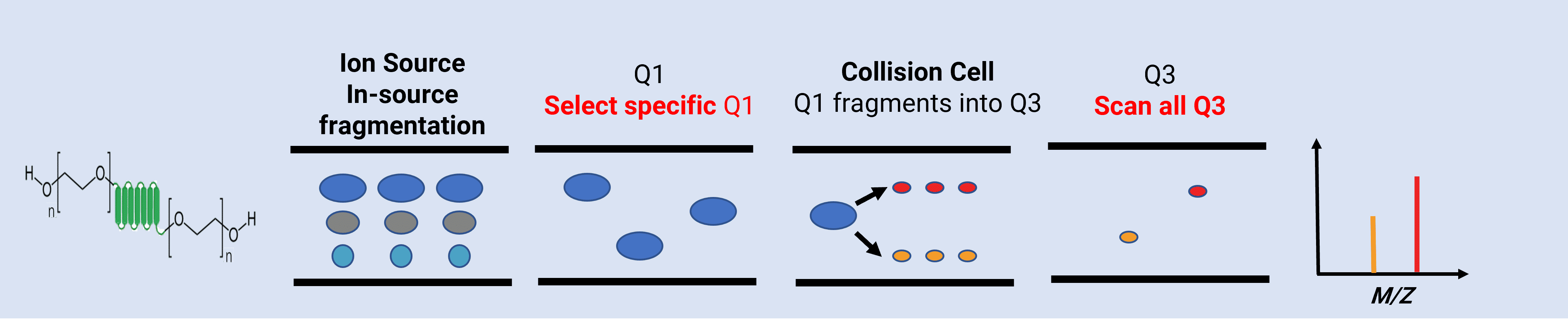
Figure 1. Schematic diagram of product ion scan
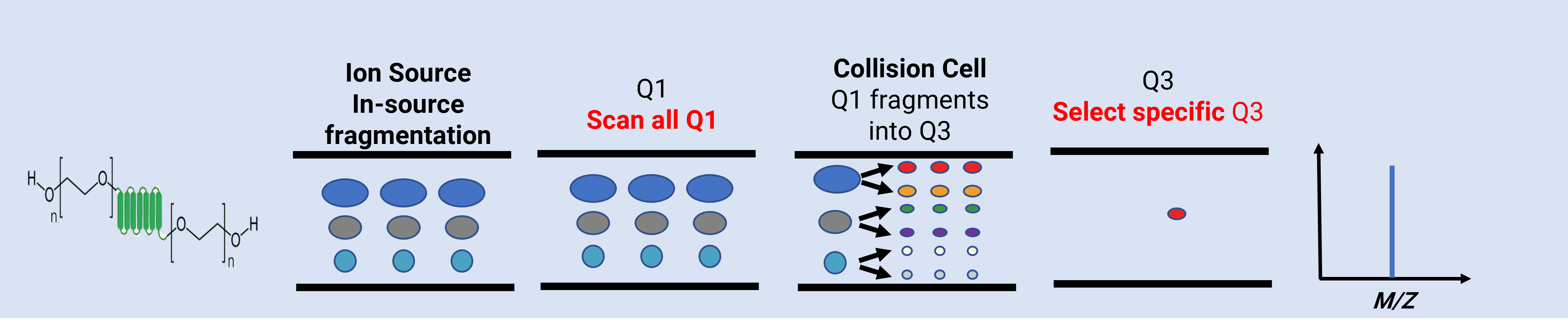
Figure 2. Schematic diagram of precursor ion scan
The core advantage of this mode is that fragments generated from the PEG chain are non-specific, whereas fragments from the peptide core ─ such as characteristic amino acid fragments and peptide fragments ─ are highly specific. Once a peptide-specific Q3 is locked, PEG interference can be avoided, and the corresponding Q1 can be precisely traced. This fundamentally resolves the difficulty of determining MRM transitions for PEGylated peptides.
Analytical Method Optimization: Coordinated Adaptation of Chromatography and Mass Spectrometry for Peptide Bioanalysis
After successful establishment of MRM transitions, chromatographic peak shape and response must be optimized to construct a complete LC-MS/MS analytical method that satisfies the requirements for quantitative accuracy and sensitivity.
Chromatographic Peak Shape Optimization
The macromolecular structure and strong hydrophilicity of PEGylated peptides often cause peak tailing and asymmetry under conventional chromatographic conditions, affecting resolution and quantitative accuracy.
Two key optimization measures are adopted to address this problem:
Selection of a large-pore chromatographic column: A large-pore column allows PEGylated peptides to interact sufficiently with the stationary phase, avoiding peak broadening caused by excessive molecular size and ensuring good resolution.
Optimization of mobile-phase additives: Conventional formic acid in the mobile phase is replaced with a protonating reagent. The protonating reagent can fully ionize H+ and protonate silanol groups on the surface of the chromatographic packing material, thereby blocking non-specific binding between PEGylated peptides and silanol groups, reducing peak tailing, and producing symmetrical and sharp chromatographic peaks.
Enhancement of Mass Spectrometric Response
Ether oxygen atoms in the PEG chain can form numerous hydrogen bonds with water molecules, producing a stable hydration layer that suppresses ionization efficiency and weakens the mass spectrometric signal response. Systematic optimization of the ion source temperature shows that temperature has a significant impact on response intensity. Increasing the temperature facilitates dissociation of the hydration layer and improves response. Combined with protein precipitation for sample pretreatment, a complete LC-MS/MS analytical method was established, achieving an LLOQ at the ng/mL level.
Conclusion
The analytical challenges of PEGylated peptides arise from their unique “peptide core + PEG chain” structure. Traditional product ion scan fails under PEG-chain interference because it follows a Q1-first strategy. By contrast, precursor ion scan adopts a Q3-first reverse strategy. By locking peptide-core-specific product ions and accurately tracing the precursor ions, it fundamentally avoids PEG-chain interference and becomes the key technology for solving this analytical problem.
The analytical strategy for PEGylated peptides can be summarized as follows:
MRM transition determination: Product ion scan may be attempted first. If no suitable transition is found, switch to precursor ion scan. At the same time, increase the declustering voltage to promote in-source fragmentation and improve the success rate.
Chromatographic optimization: Use a large-pore chromatographic column and a protonating reagent as the mobile-phase additive to eliminate peak tailing and ensure good retention time and resolution.
Mass spectrometric optimization: Increase the ion source temperature to remove the PEG hydration layer and enhance signal response. Other ion source parameters should also be adjusted to further optimize sensitivity.
As enthusiasm for the development of PEGylated peptide drugs continues to grow, the application of precursor ion scan mode will become increasingly widespread. This approach not only solves current analytical challenges, but also provides a reproducible strategy for the analysis of future PEG-modified drugs with higher molecular weights and more complex structures, helping accelerate peptide drug development.
With extensive bioanalytical experience in PEGylated peptide drugs, WuXi AppTec DMPK provides professional and efficient integrated solutions to complete peptide pharmacokinetic and metabolic transformation evaluations for this class of drugs, offering solid support for the development and successful regulatory submission of innovative drugs.
Authors: Nuoyi Zhou, Yanfeng Liu, Xinna Cui, Weimin Hu, Lili Xing
Talk to a WuXi AppTec expert today to get the support you need to achieve your drug development goals.
Committed to accelerating drug discovery and development, we offer a full range of discovery screening, preclinical development, clinical drug metabolism, and pharmacokinetic (DMPK) platforms and services. With research facilities in the United States (New Jersey) and China (Shanghai, Suzhou, Nanjing, and Nantong), 1,000+ scientists, and over fifteen years of experience in Investigational New Drug (IND) application, our DMPK team at WuXi AppTec are serving 1,700+ global clients, and have successfully supported 1,800+ IND applications.
Reference
[1] Al Shaer D, Al Musaimi O, Albericio F, de la Torre BG.2023 FDA TIDES(Peptides and Oligonnucleotides) Harvest[J]. Pharmaceuticals.2024;17(2):243.
[2] Frost & Sullivan. Global peptide therapeutics market report 2024-2030[R]. London: Frost & Sullivan,2024.
[3] Hillmen P, Szer J, Weitz I, et al. Pegcetacoplan versus eculizumab in paroxysmal nocturnal hemoglobinuria[J]. New England Journal of Medicine,2021,384(11):1028-1037.
[4] Kang J, Deluca P, Lee K. Emerging PEGylated drugs[J]. Expert Opinion on Emerging Drugs,2009,14(3):363-380.
[5]Becker, R., Dembek, C., White, L. A., & Garrison, L. P. The cost offsets and costeffectiveness associated with pegylated drugs: a review of the literature[J]. Expert Review of Pharmacoeconomics & Outcomes Research, 2012, 12(6): 775-793.
[6] Hu, X., Olivier, K., Polack, E., et al. In vivo pharmacology and toxicology evaluation of polyethylene glycol-conjugated interferon beta-1a[J]. Pharmacol. Exp. Ther., 2011,338:984-996.


Related Services and Platforms


-
 DMPK BioanalysisLearn More
-
Novel Drug Modalities DMPK Enabling PlatformsLearn More
-
Novel Drug Modalities BioanalysisLearn More
-
Small Molecules BioanalysisLearn More
-
Bioanalytical Instrument PlatformLearn More
-
PROTAC DMPK ServicesLearn More
-
ADC DMPK ServicesLearn More
-
Antibody-Oligonucleotide Conjugate (AOC) DMPK ServicesLearn More
-
Oligo DMPK ServicesLearn More
-
PDC DMPK ServicesLearn More
-
Peptide DMPK ServicesLearn More
-
mRNA DMPK ServicesLearn More
-
Covalent Drugs DMPK ServicesLearn More




Stay Connected
Keep up with the latest news and insights.

