



Decoding Peptide Metabolism: The Role of Peptide Enzymes and Peptide Optimization Strategies
-

Articles
-

Jul 23, 2026
Since insulin was used as the first peptide drug for the treatment of type 1 diabetes in 1922, peptides have entered the field of drug research and development due to their diverse physiological regulatory effects, notable activity, high selectivity, and low toxicity. Over the past decade, with the surge in biomedical R&D investment, peptide drugs have gained increased attention and development.
Peptide drugs also have non-negligible weaknesses; their chemical and physiological stability is relatively poor, and they are vulnerable to degradation by proteolytic enzymes in the body. This results in poor oral absorption, rapid peptide metabolism, short half-life, and other problems, bringing many challenges to the research and development of peptide drugs. DMPK studies can help optimize the structure of peptide drugs, evaluate the appropriate route of administration, and determine metabolism-related species, which are important processes in peptide drug research and development. In this section, the mechanism of peptide metabolism, how to carry out peptide stability testing, and how to improve peptide metabolic stability will be introduced and discussed.
What Are the Key Peptide Enzymes and Sites in Metabolism
Peptide bonds are the natural metabolic sites of proteolytic enzymes, and peptide drugs will be metabolized by proteolytic enzymes into active or inactive metabolites and excreted out of the body. There are approximately 560 human and 640 mouse proteases known, and these metabolic processes occur widely and rapidly, resulting in the short half-life of peptide drugs in vivo. Figure 1 shows the classification of proteolytic enzymes and the mode of catalysis [1].
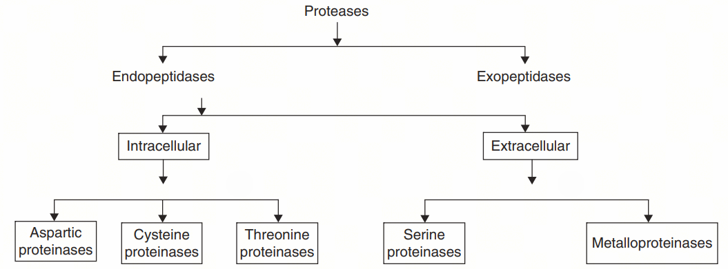
Figure 1. Main types of protease [1]
Endopeptidases break peptide bonds within the molecule. Their catalytic mechanism is related to the chemical groups involved in the hydrolysis process and can be subdivided into five different categories: aspartic proteases, cysteine proteases, threonine proteases, serine proteases, and metalloproteases. Exopeptidases specifically cleave substrates at the N- or C-terminus of peptides and can therefore be subdivided into aminopeptidases and carboxypeptidases.
The process of peptide metabolism by peptidases is non-specific and multistep. A peptidase can metabolize various types of substrates, or a substrate can be metabolized by multiple peptidases and is a sequential multistep degradation process. Peptides are usually metabolized in the body by endopeptidases to oligopeptides, which are then further degraded to amino acids by exopeptidases and subsequently involved in various biochemical processes such as protein synthesis in the body [2].
Proteases are mainly distributed in plasma, gastrointestinal tract, blood-brain barrier, liver, spleen, kidney, and other tissues, resulting in poor absorption and tissue distribution of peptide drugs and thus inconvenience of administration and inability to reach the target site and other defects. Table 1 shows the major protease categories and mode of peptide metabolism.
Table 1. Major Protease Species and Associated Amino Acid Substrates
Protease | Relevant amino acid substrates |
Pepsin | Leu, Phe, Trp, Tyr (N-terminal residues) |
Trypsin | Lys, Arg (C-terminal residues) |
Chymotrypsin | Phe, Tyr, Trp (C-terminal residues) |
Elastase | Ala, Gly, Ser, Val (C-terminal residues) |
Aminopeptidase | - |
Carboxypeptidase A, carboxypeptidase B | Phe, Tyr, Trp or Leu, Lys or Arg |
Dipeptidyl peptidase IV (DPP IV or DPP-4) | Ala, Pro |
Leu, leucine; Phe, phenylalanine; Trp, tryptophan; Tyr, tyrosine; Lys, lysine; Arg, arginine; Ala, alanine; Gly, glycine; Ser, serine; Val, valine; Pro, proline.
DPP IV is abundant in the kidney, small intestine, submaxillary gland, and liver, and some are present in the circulating blood system in a soluble form. The activity is optimal at a pH of approximately 7.8 and maintains similar activity even over a wide range of pH 5 to 10. DPP IV is very sensitive to the protease inhibitor diisopropyl fluorophosphate (DFP), but much less sensitive to other common serine protease inhibitors, such as diethyl p-nitrophenyl phosphate and phenylmethanesulfonyl fluoride (PMSF) [3].
Glucagon-like peptide-1 (GLP-1) is an important hormone that promotes insulin secretion and controls blood glucose. It is rapidly degraded by DPP IV in vivo, with a half-life of 0.9 min in humans [4]. GLP-1 receptor agonists such as exenatide, liraglutide, dulaglutide, and lixisenatide have effects similar to native GLP-1, and are used to treat type 2 diabetes and manage weight. This is because structural optimization reduces degradation caused by DPP IV and prolongs the duration of action. DPP IV inhibitors such as sitagliptin are also used as drugs for the treatment of diabetes. Figure 2 shows the mechanism of action of DPP IV inhibitors.
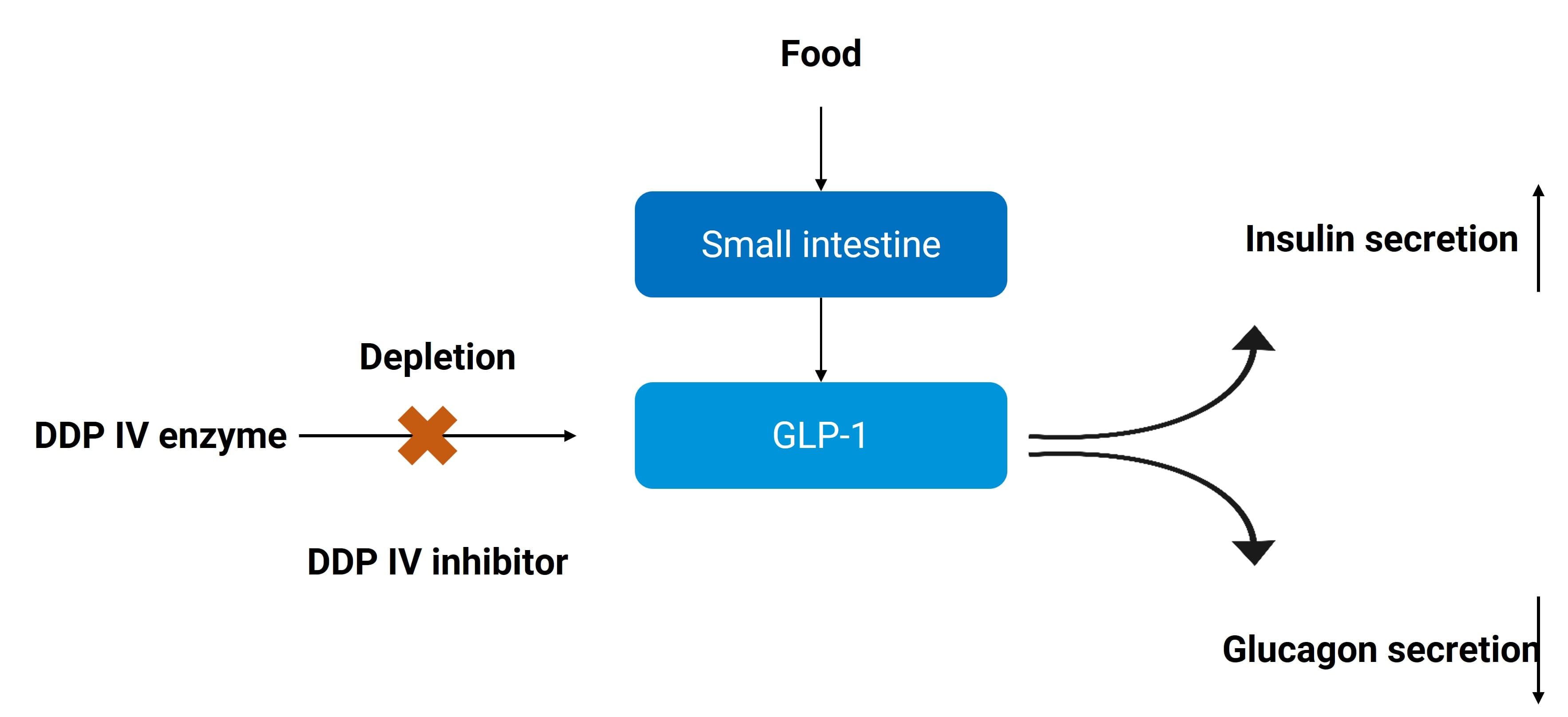
Figure 2. Mechanism of Action of DPP IV Inhibitors[23]
Gastrointestinal metabolism
The gastrointestinal tract contains abundant proteases, which become the main barrier to peptide absorption. Figure 3 shows the secretion and activation of proteases in the gastrointestinal tract. There are three sources of these proteases:
Secreted enzymes, including pepsin, trypsin, chymotrypsin, elastase, carboxypeptidase A, and carboxypeptidase B.
Brush border membrane-bound enzymes, including various carboxypeptidases and aminopeptidases.
Proteases and other oxidases in the cytoplasm of gastrointestinal epithelial cells.
The stability of 17 kinds of peptide drugs in the gastrointestinal tract was studied in a paper [5], and the peptide drugs were divided into three classes according to the number of amino acids.
Five drugs with less than 12 amino acids and with partial or all cyclic structures.
Seven linear peptide drugs of less than 12 amino acids.
Five drugs greater than 12 amino acids.
The findings suggest that in human gastric fluid, larger peptides including somatostatin, calcitonin, secretin, glucagon, and insulin are metabolized rapidly, whereas smaller ones showed good stability. However, in human small intestinal fluid, both smaller and larger peptides are rapidly degraded except for cyclic peptide cyclosporin and disulfide-bridged peptide octreotide and desmopressin. The peptide metabolic stability in simulated and porcine gastric fluid correlated well with the stability in human gastric fluid. The rapid degradation of larger polypeptides in gastric fluid may be due to multiple factors. For example, larger peptides have a large number of pepsin-sensitive peptide bonds, are structurally flexible, have more hydrogen bond acceptors/donors, and have a higher polar surface area, thereby increasing the interaction with pepsin.
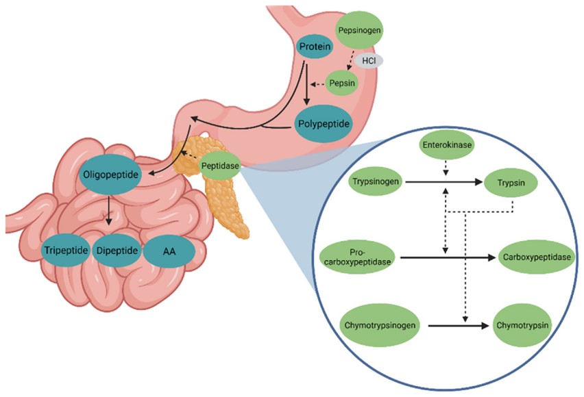
Figure 3. Secretion and Activation of Proteases in the Gastrointestinal Tract [6]
Hepatic metabolism
Peptide drugs are mostly less hydrophobic and cannot enter hepatocytes by passive diffusion. They will be metabolized after entering hepatocytes via carrier-mediated transmembrane transport or endocytosis. Receptors associated with low-density lipoprotein (LDL) receptors found on hepatocyte membranes may be involved in the metabolism of plasminogen activators [7]. A study on the metabolism of two GLP-1 metabolites in human and mouse hepatocytes showed that both metabolites are rapidly metabolized at the N-terminus, but not at the C-terminus [8]. Salmon calcitonin is another peptide metabolized by the liver, and when incubated in vitro with rat liver homogenates, salmon calcitonin is first cleaved at the His17-Lys18 and Val8-Leu9 bonds, and then these cleavage products are further degraded by exopeptidases (aminopeptidase/carboxypeptidase) [9].
CYP450 enzymes in the liver can also metabolize a variety of polypeptide drugs. For example, the prodrug of the opioid peptide H-Tyrd-AlaGlyPhed-LeuOH (DADLE) is sensitive to CYP450 oxidation [10].
Renal Metabolism
Depending on the molecular weights, Polypeptides are metabolized in the kidney by two main mechanisms. After glomerular filtration, macromolecular peptides will be removed by endocytosis and lysosomal degradation and finally hydrolyzed into small peptides and amino acids. Peptides with smaller molecular weights will be hydrolyzed to amino acids by exopeptidase on the brush-border membrane of the tubule after glomerular filtration, and then reabsorbed into the systemic circulation or degraded to small peptides and transported to the proximal tubule epithelial cells through specific amino acid transport systems. Peptide transporter 1 (PEPT1) and PEPT2 are involved in the reabsorption mechanism of oligopeptides.
After subcutaneous injection of teriparatide acetate in rats, kinetic data show that the kidney is the main organ of its distribution and degradation, but excretion is not significant [11]. The metabolism of natriuretic peptides (NPs) serves as another example. D-type NPs (DNPs) are more stable in rabbit plasma compared with other natriuretic peptides (e.g., atrial natriuretic peptides); Further investigating the metabolism of DNP in other organs and the results indicate that the kidney is the main organ for its degradation [12].
Blood Metabolism
Peptide drugs are hydrophilic and are more likely to be degraded by proteases in plasma than in tissues and organs. For example, bradykinin is rapidly degraded by proteases found in human plasma [13]. Neuropeptide Y (NPY) is also metabolized by proteolytic cleavage when incubated in human serum. NPY1-36 can be rapidly converted to three metabolites, and this degradation process can be prevented by inhibiting DPP IV, aminopeptidase P, and kinin-releasing enzyme [14].
Metabolism in other tissues
Because of the poor oral bioavailability of peptide drugs, the most common route of administration in clinical practice is intravenous or subcutaneous administration. When administered subcutaneously, the drug is also metabolized in the subcutaneous tissue before entering the systemic circulation. Subcutaneous interstitial fluid (SIF) contains proteases similar to plasma, but the content is only one-third of plasma, similar to lymph [15]. The albumin content of subcutaneous tissue is less than that of plasma. For the polypeptide bound to albumin to prolong the half-life, the free ratio in subcutaneous tissue is higher than that in plasma, therefore, subcutaneous tissue is its main metabolic site. Ito et al. [16] incubated leuprorelin in rat skin tissue samples to account for the low subcutaneous bioavailability observed in vivo.
Receptor-mediated metabolism
Because peptide drugs have better target affinity, a large proportion of drug molecules will bind to target receptors. Peptide drugs are usually internalized into cells after binding to target receptors on the cell membrane surface and degraded by intracellular endosomes or lysosomes. Due to the limited number of target receptors, this route of elimination is easily saturated. Therefore, this pathway is one of the important reasons for the nonlinear PK properties of peptides or macromolecular drugs. Target receptors expressed in the blood generally have a more significant effect on the elimination of peptide drugs than target receptors expressed in tissue cells.
How to Conduct Peptide Stability Testing and Identify Metabolic Sites
As mentioned above, peptides are easily metabolized by proteases in tissues such as blood, liver, kidney, gastrointestinal tract, and skin, so in vitro incubation at 37°C in blood or plasma is often used to investigate stability. In addition, liver S9, kidney S9, simulated gastrointestinal fluid, and skin homogenate are also recommended peptide stability test systems. It should be noted that because of the low permeability of peptides, in vitro S9, homogenate, and other systems may overestimate the extent of peptide metabolism in tissues in vivo. Metabolites of peptides are usually also biologically active, and high-resolution mass spectrometry (HRMS) can be used to study the metabolic sites and products of peptides in preclinical practice to help guide structure optimization to improve peptide metabolic stability and focus on the activity and toxicity of major metabolites.
If the main types of metabolic enzymes of drugs are understood, stability testing using recombinant proteases will provide more direct results. However, there are few types of proteases commercially available, and specific protease inhibitors can also be considered to investigate the metabolic phenotype.
How to Achieve Peptide Optimization and Improve Peptide Metabolic Stability
In response to the problems of rapid peptide metabolism and short duration of action, medicinal chemists have explored a variety of optimization methods to improve peptide metabolic stability.
Substitution of L-Amino Acids for D-amino Acids or Unnatural Amino Acids
The primary recognition site for protease in organisms is the natural L-amino acids. Thus, substituting L-amino acids with D-amino acids at the metabolic site can reduce the affinity towards protease while preserving the physiological activity of the polypeptide.
Triptorelin (10-amino acid peptide), whose structure is shown in Figure 4, is a gonadotropin-releasing hormone (GnRH) analog. The amino acid residues at positions 1, 6, and 10 of native GnRH are substituted with three unnatural amino acid residues, and its half-life after intravenous injection is prolonged to 2.8 hours compared with 5 minutes of native GnRH, which is mainly cleared by the liver and kidney [17].
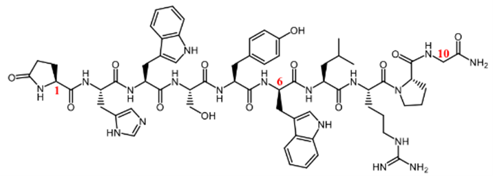
Figure 4. Structural formula of triptorelin (unnatural amino acid residues in red) [17]
N- or C-Terminal Modification
Amino acid residues at both ends of linear peptides are susceptible to degradation by exopeptidases, so modification of terminal residues by methylation or N-acetylation can play a role in metabolic protection.
The half-life of the glucose-dependent insulinotropic polypeptide (GIP) (1-42) is only 2 to 5 min, and Mabilleau et al. developed an enzymatically stable GIP analogue by acetylating Tyr1 (N-AcGIP) in GIP with an in vivo half-life of greater than 24 h.
Cyclization or Bicyclization
The configuration of the linear peptide is unstable and easily transformed to bind to the protease catalytic center. The method of cyclization can stabilize the peptide configuration, reduce the binding constant (Ka) to the protease, and reduce the affinity to the protease. It can also protect the amino acid residues at both ends or metabolic sites, which is an effective way to enhance peptide metabolic stability. At present, more than two-thirds of the peptide drugs that have been marketed are cyclic peptide compounds [18].
Many antimicrobial agents from natural sources are cyclic peptides, such as cyclosporin and daptomycin. Cyclosporin possesses high metabolic stability against gastrointestinal peptidases (> 90% remains intact after 2 h of incubation) [19]. Sandimmune ® is one of its lipid-based formulations, while Neoral ® is a microemulsified formulation with improved bioavailability. Its terminal half-life ranges from 5 to 18 hours after oral administration.
Lanreotide is an 8-residue cyclic peptide analogue of 14-amino acid peptide somatostatin for the treatment of acromegaly (gigantism) and unresectable advanced or metastatic gastroenteropancreatic neuroendocrine tumors (GEP-NETs) caused by excessive hormone release from the pituitary gland. The investigators developed a 1-month sustained release subcutaneous depot formulation (Somatuline Autogel ® and Somatuline Depot ®) to avoid daily injections. Lanreotide contains two unnatural amino acid residues at positions 1 and 4, followed by amidated Thr8, extending its elimination half-life to 23 to 30 days, compared with 3 min for endogenous somatostatin [20].
PEGylation
PEG has a variety of drug-related properties: high water solubility, high fluidity, low toxicity, and immunogenicity, and is easily removed from the body. When the drug is PEGylated, it acquires certain properties, and the transmission of these properties positively correlates with the molecular weight of PEG. PEGylation modification at the end of the peptide chain not only protects the amino acid residues, but also significantly increases the molecular weight and steric hindrance. Research by Lee et al. demonstrated that site-specific PEGylation of GLP-1 resulted in a 16-fold increase in plasma half-life in rats [21].
Another significant function of PEGylation with large molecular weight is to decrease glomerular filtration rate and reduce renal clearance. The combination of both mechanisms can greatly prolong the elimination half-life in vivo of peptides.
Addition of Ligand Improves Binding to Albumin or IgG
Albumin or IgG in the organism uses the neonatal Fc receptor (FcRn) cycle (Figure 5) pathway to avoid degradation. When albumin is internalized into cells, it binds to FcRn at pH 6 in the endosome, protecting albumin from lysosomal degradation. The conjugates are then released into the extracellular environment at physiological pH 7.4 to release albumin. In relation to this pathway, researchers have modified the peptide structure with the aliphatic chain to bind albumin, and both liraglutide and semaglutide have significantly extended the half-life in this way. Other research approaches also include covalent binding of peptides to albumin and recombinant expression [22].
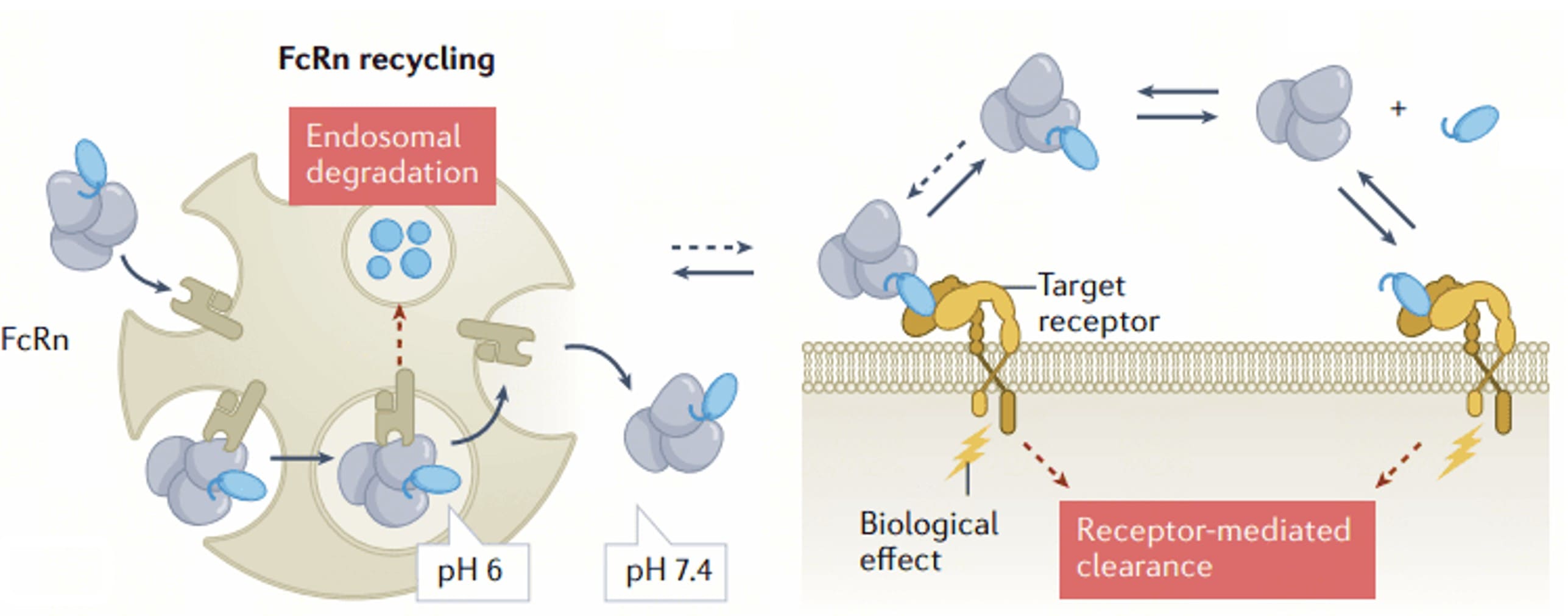
Figure 5. FcRn Cycle [22]
In addition to the above ways, the in vivo half-life of the peptide drug can also be prolonged by co-administration with peptidase enzyme inhibitors and by using sustained-release formulations such as liposomes to protect free peptides.
Final Words
Deepening the understanding of peptide metabolic stability and early identification of metabolic mechanisms and soft spots can aid researchers in conducting structural modifications to improve drug stability, which is crucial for successful clinical development. Nowadays, several long-acting peptide drugs and sustained-release delivery formulations have successfully entered the market. With the advancement of more research, including the exploration of new molecular types such as multifunctional peptides and peptide-drug conjugates, peptides stand a chance of being utilized in a wider array of disease areas.
Authors: Jianping Sun, Jing Jin
Talk to a WuXi AppTec expert today to get the support you need to achieve your drug development goals.
Committed to accelerating drug discovery and development, we offer a full range of discovery screening, preclinical development, clinical drug metabolism, and pharmacokinetic (DMPK) platforms and services. With research facilities in the United States (New Jersey) and China (Shanghai, Suzhou, Nanjing, and Nantong), 1,000+ scientists, and over fifteen years of experience in Investigational New Drug (IND) application, our DMPK team at WuXi AppTec are serving 1,600+ global clients, and have successfully supported 1,800+ IND applications.
Reference
[1] Pernot, M., Vanderesse, R., Frochot, C. et al. (2011). Stability of peptides and therapeutic success in cancer. Expert Opinion Drug Metabolism and Toxicology 7: 793-802.
[2] Yao, J.F., Yang, H., Zhao, Y.Z., and Xue, M. (2018). Metabolism of peptide drugs and strategies to improve their metabolic stability. Current Drug Metabolism 19 (11): 892-901.
[3] Rawlings, N.D. and Salvesen, G. (ed.) (2013). Handbook of Proteolytic Enzymes, 3e. Amsterdam: Elsevier/AP.
[4] Zhu, L., Tamvakopoulos, C., Xie, D. et al. (2003). The role of dipeptidyl peptidase IV in the cleavage of glucagon family peptides in vivo metabolism of pituitary adenylate cyclase-activating polypeptide- (1-38). Journal of Biological Chemistry 278: 22418-22423.
[5] Wang, J., Yadav, V., Smart, A.L. et al. (2015). Toward oral delivery of biopharmaceuticals: an assessment of the gastrointestinal stability of 17 peptide drugs. Molecular Pharmaceutics Journal 12 (3): 966-973.
[6] Kurz, A. and Seifert, J. (2021). Factors influencing proteolysis and protein utilization in the intestine of pigs: a review. Animals 11 (12): 3551. http: //doi.org/10.3390/ani11123551.
[7] Authier, F., Posner, B.I., and Bergeron, J.J.M. (1996). Endosomal proteolysis of internalized proteins. FEBS Letters 389: 55-60.
[8] Sharma, R., McDonald, T.S., Eng, H. et al. (2013). In vitro metabolism of the glucagon-like peptide-1 (GLP-1) -derived metabolites GLP-1 (9-36) amide and GLP-1 (28-36) amide in mouse and human hepatocytes. Drug Metabolism & Disposition 41: 2148-2157.
[9] Liao, S., Qie, J.K., Xue, M. et al. (2010). Metabolic stability of human parathyroid hormone peptide hPTH (1-34) in rat tissue homogenates: kinetics and products of proteolytic degradation. Amino Acids 38: 1595-1605.
[10] Nofsinger, R., Fuchs-Knotts, T., Borchardt, R.T. (2012). Factors that restrict the cell permeation of cyclic prodrugs of an opioid peptide, part 3: synthesis of analogs designed to have improved stability to oxidative metabolism. Journal of Pharmacological Sciences 101: 3486-3499.
[11] Serada, M., Sakurai-Tanikawa, A., Igarashi, M. et al. (2012). The role of the liver and kidneys in the pharmacokinetics of subcutaneously administered teriparatide acetate in rats. Xenobiotica 42 (4): 398-407.
[12] Kim, S.M., Kim, S.Y., Kim, S.H., and Kim, S.Z. (2014). Dendroaspis natriuretic peptide is degraded by a metalloproteinase in the rat kidney. Molecular Medicine Report 9: 1037-1043.
[13] Laine, J., Murphey, D.L., Hachey, J.A. et al. (2000). Metabolism of bradykinin in vivo in humans: Identification of BK1-5 as a stable plasma peptide metabolite. Journal of Pharmacology and Experimental Therapeutics 294: 263-269.
[14] Abid, K., Rochat, B., Lassahn, P.G. et al. (2009). Kinetic study of neuropeptide Y (NPY) proteolysis in blood and identification of NPY3-35 a new peptide generated by plasma kallikrein. Journal of Biological Chemistry 284: 24715-24724.
[15] Esposito, S., Orsatti, L., and Pucci, V. (2022). Subcutaneous catabolism of peptide therapeutics: bioanalytical approaches and ADME considerations. Xenobiotica 52 (8): 828-839. http://doi.org/10.1080/00498254.2022.2119180.
[16] Ito, Y., Murano, H., Hamasaki, N. et al. (2011). Incidence of low bioavailability of leuprolide acetate after percutaneous administration to rats by dissolving microneedles. International Journal of Pharmaceutics 407 (1-2): 126-131.
[17] Muller, F.O., Terblanchè, J., Schall, R. et al. (1997). Pharmacokinetics of triptorelin after intravenous bolus administration in healthy and in males with renal or hepatic insufficiency. British Journal of Clinical Pharmacology 44 (4): 335-341.
[18] Zorzi, A., Deyle, K., and Heinis, C. (2017). Cyclic peptide therapeutics: past, present and future. Current Opinion of Chemical Biology 38: 24-29.
[19] Wang, J., Yadav, V., Smart, A.L. et al. (2015). Toward oral delivery of biopharmaceuticals: an assessment of the gastrointestinal stability of 17 peptide drugs. Molecular Pharmaceutics Journal 12 (3): 966-973.
[20] Brian Chia, C.S. (2021). A Review on the metabolism of 25 peptide drugs. International Journal of Peptide Research and Therapeutics 27: 1397-1418. http://doi.org/10.1007/s10989-021-10177-0.
[21] Lee, S.H., Lee, S., Youn, Y.S. et al. (2005). Synthesis, characterization, and pharmacokinetic studies of PEGylated glucagon-like peptide-1. Bioconjugate Chemistry 16:377-382.
[22] Kurtzhals, P., Østergaard, S., Nishimura, E. et al. (2022). Derivatization with fatty acids in peptide and protein drug discovery. Nature Reviews Drug Discovery. 22 (1): 59-80.
[23] Liang Shen, editor. Drug Metabolism and Pharmacokinetics: Frontiers, Strategies, and Applications. Wiley. 2025. http://doi.org/10.1002/9781394300150.


Related Services and Platforms


-
 MetID (Metabolite Profiling and Identification)Learn More
-
Novel Drug Modalities DMPK Enabling PlatformsLearn More
-
In Vitro MetID (Metabolite Profiling and Identification)Learn More
-
In Vivo MetID (Metabolite Profiling and Identification)Learn More
-
Metabolite Biosynthesis and Structural CharacterizationLearn More
-
Metabolites in Safety Testing (MIST)Learn More
-
PROTAC DMPK ServicesLearn More
-
ADC DMPK ServicesLearn More
-
Antibody-Oligonucleotide Conjugate (AOC) DMPK ServicesLearn More
-
Oligo DMPK ServicesLearn More
-
PDC DMPK ServicesLearn More
-
Peptide DMPK ServicesLearn More
-
mRNA DMPK ServicesLearn More
-
Covalent Drugs DMPK ServicesLearn More


Stay Connected
Keep up with the latest news and insights.

