



Unraveling Peptide Disulfide Bonds via Enzymatic Digestion: A Rapid and Precise Characterization Strategy
-

Articles
-

Jul 31, 2026
What Is the Role of Disulfide Bonds in Peptide Therapeutics
Nearly a century has passed since the advent of insulin, the first peptide drug. To date, approximately 120 peptide drugs have been approved and launched globally. The landscape of peptide drug development continues to expand rapidly, demonstrating broad clinical applicability across indications such as diabetes, oncology, osteoporosis, multiple sclerosis, HIV, and chronic pain management. According to industry projections, the global peptide drug market is expected to surge from USD 62.8 billion in 2020 to USD 96.0 billion by 2025 [1].
However, early endogenous peptide hormones were often hindered by inherent limitations, most notably their short in vivo half-lives. To enhance the stability, potency, and target selectivity of these endogenous ligands, chemical modification has emerged as a cornerstone strategy. Common structural modifications include N-terminal acetylation, N-methylation, N- and C-terminal cyclization, PEGylation, and the incorporation of disulfide bonds [2].
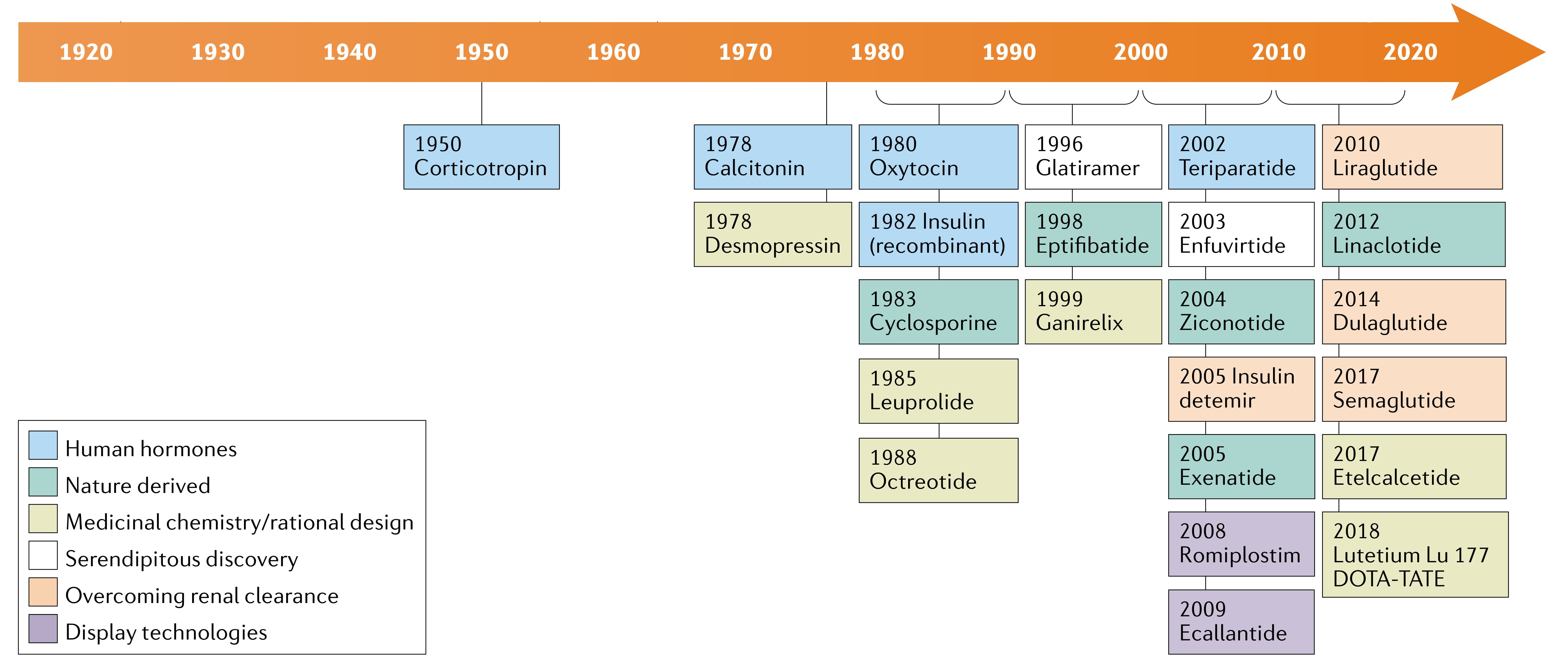
Figure 1. Development history and milestones of peptide drugs [2]
A disulfide bond is a covalent linkage formed by the oxidation of the thiol groups (-SH) on the side chains of two cysteine residues (cysteine). These bonds can bridge different peptide chains (interchain) or connect disparate regions within a single peptide chain (intrachain), as illustrated in Figure 2. In peptide therapeutics, disulfide bonds are vital for stabilizing spatial conformation, maintaining correct folding, regulating biological activity, and ensuring intended physiological functions [3].
Despite their importance, predicting the exact linkage sites of disulfide bonds directly from an amino acid sequence is notoriously difficult, particularly for complex structures harboring three or more cysteine residues. In addition, one-step random cyclization is commonly used during synthesis, further increasing uncertainty in the linkage sites. Nevertheless, detailed information on disulfide bond sites is crucial for protein function studies and for quality assessment in biopharmaceutics [4].
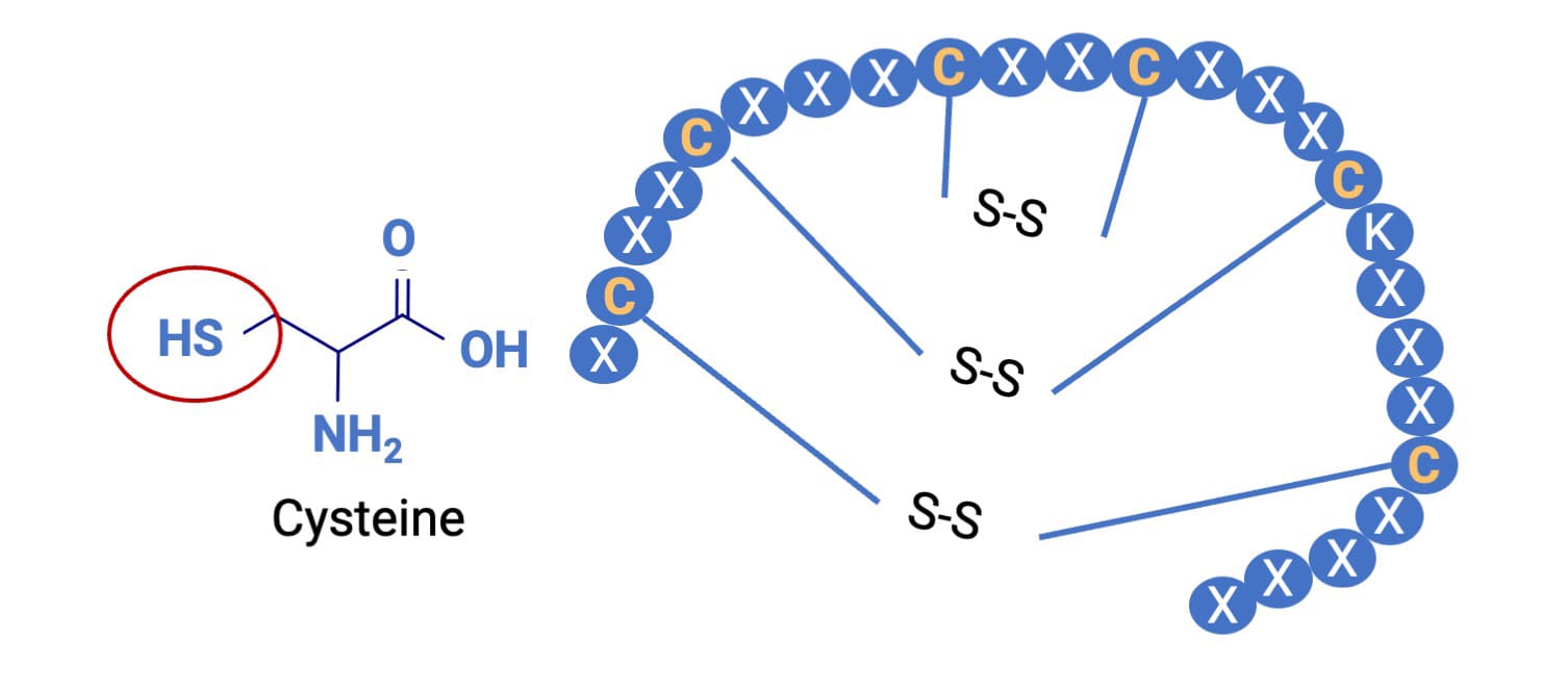
Figure 2. Chemical structures of cysteine and a disulfide bond
Historically, existing methods for disulfide bond analysis have been limited by narrow applicability and protracted turnaround times. Traditional workflows often necessitate the de novo synthesis of multiple peptide variants followed by labor-intensive comparative testing. Consequently, there is an urgent industry demand for a robust, versatile, and high-throughput analytical solution. This article outlines an optimized enzymatic digestion approach that combines specific and non-specific digestion, together with the in-house developed software PeptideID, to provide a rapid and convenient route for disulfide bond characterization.
What Are the Common Methods for Disulfide Bond Analysis in Peptides
Current mainstream techniques for mapping disulfide linkages include enzymatic digestion, tandem mass spectrometry (MS/MS) fragmentation, partial reduction and alkylation, nuclear magnetic resonance (NMR), and X-ray crystallography. The fundamental principles and characteristics of these modalities are summarized below.
Enzymatic Digestion [5]
Specific hydrolases, such as trypsin, chymotrypsin, and pepsin, are deployed to cleave the peptide backbone. By isolating and analyzing the resulting digestion products in which the disulfide bonds remain intact, scientists can accurately deduce the original disulfide linkage sites (Figure 3).
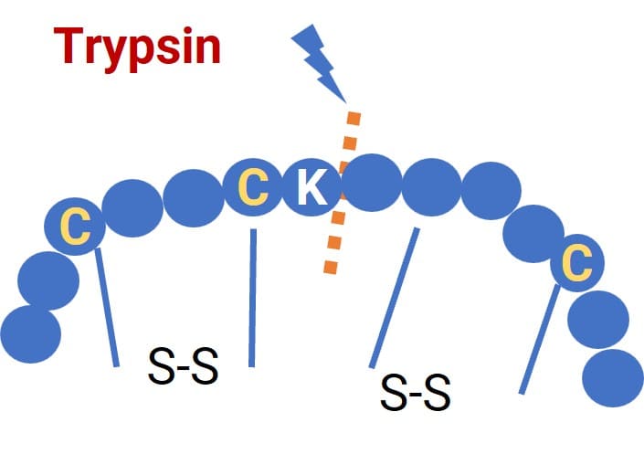
Figure 3. Example of peptide hydrolysis via enzymatic digestion
Mass Spectrometry Fragmentation [6]
MS fragmentation generates rich sets of fragment ions, allowing analysts to pinpoint diagnostic fragments that confirm specific disulfide linkages. Common fragmentation modes include:
Higher-energy collisional dissociation (HCD): Induces amide bond cleavage (b/y ions), with the potential for concurrent cleavage of disulfide bonds.
Collision-induced dissociation (CID): Primarily drives amide bond cleavage (b/y ions) while generally leaving disulfide bonds intact.
Electron-transfer dissociation (ETD): Induces amide bond cleavage (c/z ions) accompanied by preferential cleavage of the disulfide bonds.
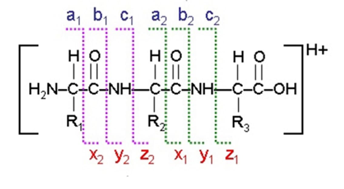
Figure 4. Classification of fragment ions in mass spectrometric fragmentation [6]
Partial Reduction
Reducing agents such as TCEP and DTT, or photochemical reduction, can be utilized to partially reduce disulfide bonds. In combination with derivatization to cap free thiol groups, mass spectrometric analysis of the structural shifts before and after reduction can reveal the linkage sites.
For instance, utilizing post-column reduction (Figure 5), interchain disulfide bonds are partially reduced during liquid chromatography-mass spectrometry (LC-MS) runs. By comparing the retention times and MS signals of the products pre- and post-reduction, the original disulfide positions can be confidently identified [7].
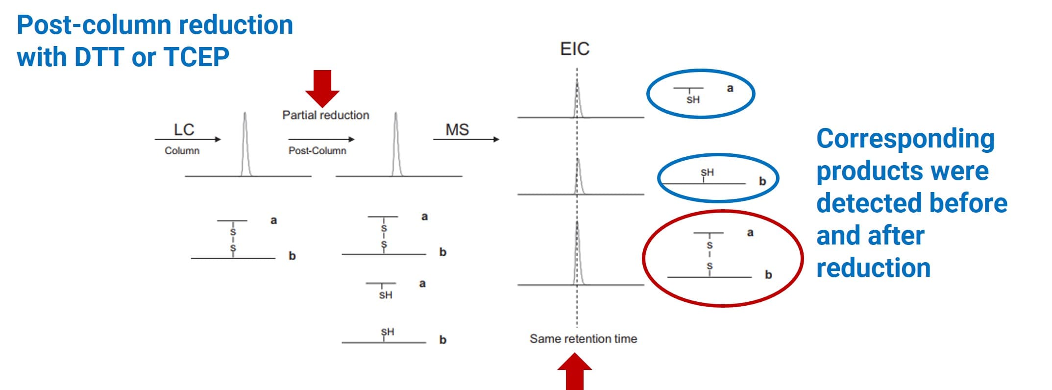
Figure 5. Example of disulfide bond characterization via post-column reduction [7]
While post-column reduction is highly effective for interchain bonds, a more versatile approach involves partial reduction followed by immediate derivatization to trap the free thiols. Subsequent MS analysis then maps the original architecture. For example, disulfide bonds can be partially photoreduced and then alkylated through a thiol-ene reaction pathway; subsequent analysis can be used to infer the original disulfide linkage sites [8].
NMR [9]
By evaluating the nuclear spin behavior of proteins within a strong magnetic field, NMR spectroscopy can resolve three-dimensional spatial structures in solution, thereby identifying disulfide bond coordinates.
X-ray Crystallography [10]
X-ray crystallography relies on protein crystal diffraction patterns to achieve atomic-level 3D structural resolution, enabling the direct visual confirmation of disulfide linkage sites.
The advantages and limitations of these analytical methodologies are compared in Table 1. When factoring in operational efficiency and throughput, enzymatic digestion stands out for its versatility and straightforward workflow, making it the method of choice for the ensuing experimental designs.
Table 1. Comparison of Advantages and Limitations of Disulfide Bond Analysis Methods
Method | Advantages | Limitations |
Enzymatic digestion | Straightforward workflow | Dependent on the presence of specific cleavage sites; digestion conditions require optimization; short peptides may exhibit poor chromatographic retention |
Mass Spectrometry Fragmentation | Straightforward workflow | Unstable dissociation performance for complex structures |
Partial Reduction | Versatile across peptide classes | Reduction conditions require optimization; applicability may differ across peptides |
NMR | Non-destructive; broad applicability | High requirements for sample purity and concentration; difficult analysis of complex molecules; long data acquisition time |
X-ray crystallography | Atomic-level structural resolution | Requires crystal cultivation; reflects only the crystalline state; high technical threshold; time-consuming |
How to Perform Disulfide Bond Mapping via Enzymatic Digestion
Capitalizing on the inherent strengths of enzymatic digestion, a dual-approach experimental framework was designed, supported by a specialized data processing workflow.
Dual-Approach Experimental Framework
Approach 1: Specific Enzymatic Digestion
This method employs proteases that recognize highly specific amino acid sequences (e.g., trypsin). It yields well-defined, predictable products, allowing for straightforward spectral interpretation. However, its utility is strictly dictated by the peptide sequence; it is rendered ineffective if the peptide lacks suitable enzymatic cleavage sites.
Approach 2: Non-Specific Enzymatic Digestion
This strategy utilizes non-specific proteases (e.g., pronase) or relies on high enzyme concentrations coupled with prolonged incubation periods to generate a complex mixture of short, overlapping peptides that cover the entire sequence. While this approach boasts near-universal applicability, digestion conditions require rigorous optimization, yielding highly complex product mixtures.
To strike an optimal balance between digestion efficiency and product characterization coverage, it is highly recommended to sample the reaction at multiple incubation time points. This dynamic sampling ensures the successful capture of transient diagnostic disulfide-linked peptides.
Methodology and Analytical Challenges
The fundamental principle of disulfide bond characterization via enzymatic digestion hinges on identifying "diagnostic fragments"—specifically, peptide fragments that retain an intact disulfide bond post-digestion and contain two or more cysteine residues (Figure 6). However, rapidly and accurately isolating these diagnostic fragments from a chaotic background of digestion products presents a significant analytical hurdle.
Primary challenges in this workflow include:
Under-digestion: If cleavage efficiency is too low, the enzyme may fail to cleave the backbone between adjacent cysteine residues, yielding fragments that are too large to be informative.
Over-digestion: Excessive digestion can induce disulfide bond cleavage or scrambling (disulfide exchange), introducing false positives and preventing accurate site localization.
Data Bottlenecks: The sheer volume of MS data generated makes manual screening prohibitively time-consuming and labor-intensive.
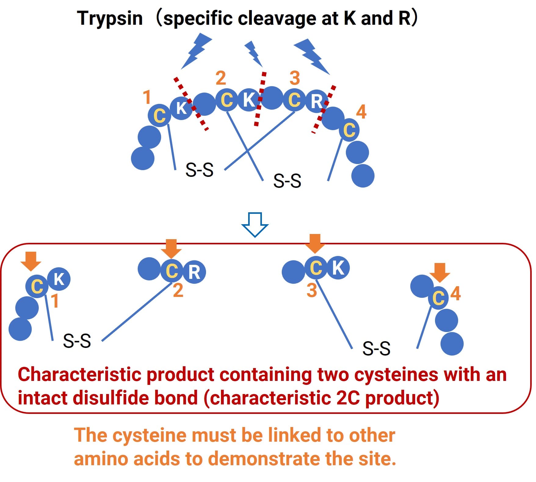
Figure 6. Diagnostic products retaining disulfide bonds after enzymatic digestion
Data Processing and Linkage Site Inference
Consider a complex peptide containing three cysteine residues, presenting three theoretical disulfide linkage permutations. The optimized experimental workflow proceeds as follows:
Execute specific enzymatic digestion, aggressively sampling the reaction at three distinct incubation time points;
Deploy the proprietary PeptideID software to rapidly and automatically screen for diagnostic products corresponding to each theoretical linkage permutation;
Import the curated diagnostic product list into Compound Discoverer software to screen and match against the raw empirical LC-MS data;
Deduce the most probable disulfide linkage sites based on the specific types and relative abundances of the successfully matched diagnostic products (Figure 7).
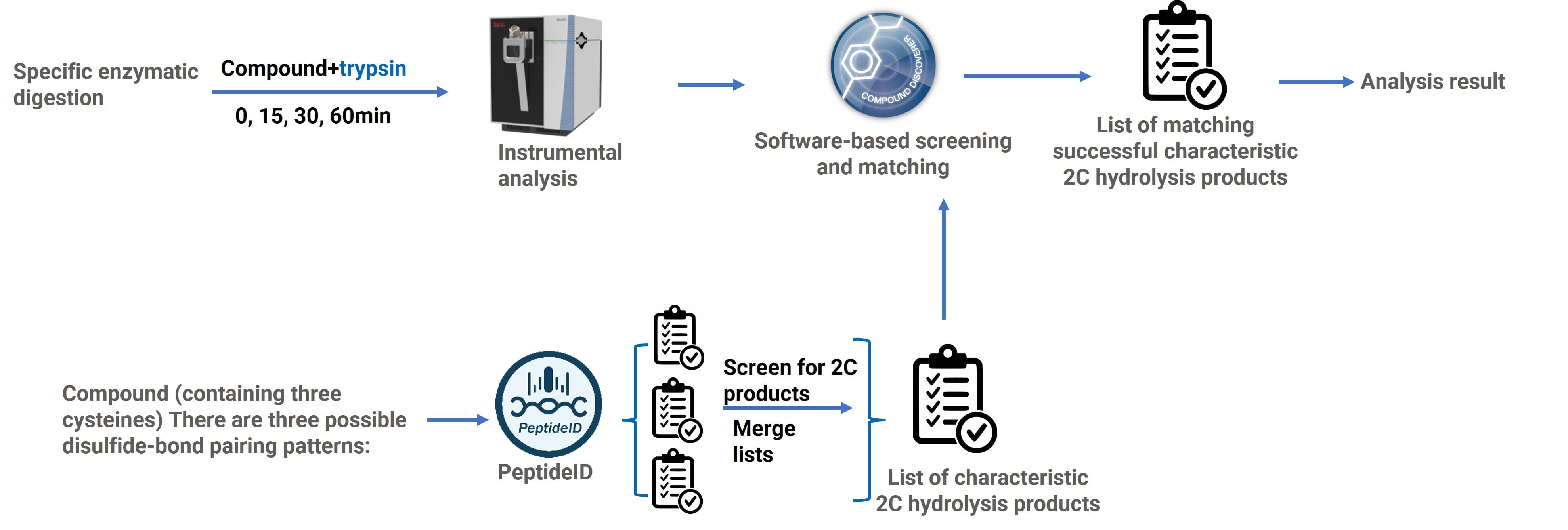
Figure 7. Data processing workflow for disulfide bond mapping by enzymatic digestion.
Comparison of Specific and Non-specific Enzymatic Digestion Methods
The scope of application and inherent limitations of both digestion strategies are outlined in Table 2.
Table 2. Comparison of Specific and Non-Specific Enzymatic Digestion Strategies
Experimental Strategy | Scope of Application | Limitations |
Specific enzymatic digestion | The sequence contains clearly defined enzymatic cleavage sites | Highly sequence-dependent; enzymatic activity conditions require optimization |
Non-specific enzymatic digestion | Applicable to the vast majority of peptide structures | Digestion products are difficult to control; extensive condition screening is required |
For complex cyclic peptides containing three or more cysteine residues, WuXi AppTec DMPK has established a robust, tiered decision tree (Figure 8):
Prioritize specific enzymatic digestion: If specific enzymatic cleavage sites are present in the sequence, specific enzymatic digestion should be performed first. If diagnostic products can be successfully matched, the disulfide linkage sites can be directly inferred.
Apply non-specific enzymatic digestion: If specific digestion cannot yield a clear conclusion, or if the sequence itself lacks suitable cleavage sites, non-specific enzymatic digestion should be performed. Disulfide linkage sites are inferred through multi-time-point sampling, high-resolution mass spectrometric detection, and diagnostic product matching.
Alternative approaches: If neither digestion strategy can clearly determine the disulfide linkage sites, other methods such as reduction-based methods, mass spectrometric fragmentation, or NMR should be introduced for integrated analysis.
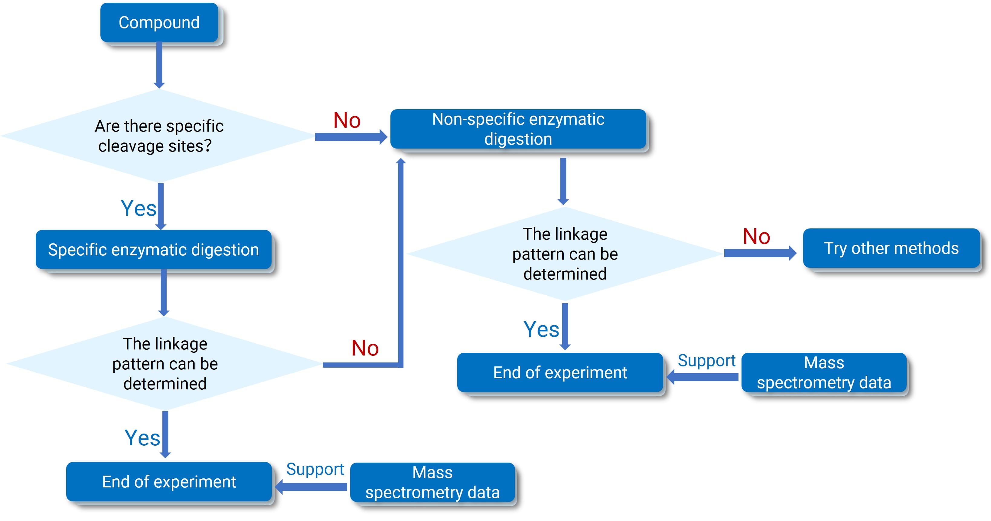
Figure 8. Decision tree for peptide disulfide bond characterization
Conclusion
The momentum behind peptide drug development continues to accelerate across the global biopharmaceutical landscape. Within this context, disulfide bond analysis remains a technically demanding yet indispensable step for structural confirmation and regulatory quality control. Drawing upon extensive bioanalytical project experience, WuXi AppTec DMPK has successfully engineered an accurate, rapid, and universally applicable workflow to conquer this analytical challenge.
This advanced strategy innovatively merges specific and non-specific enzymatic digestion techniques. By capturing critical diagnostic fragments via dynamic multi-time-point sampling, and pairing this empirical data with the intelligent screening capabilities of our proprietary PeptideID software, this workflow dramatically accelerates data interpretation and guarantees the precise localization of disulfide bonds in highly complex peptides.
To date, this methodology has been successfully deployed across numerous client projects, resolving diverse structural architectures containing between three and six cysteine residues. Proven in practice, this integrated workflow rapidly clarifies disulfide linkage patterns during early-stage discovery, significantly mitigating trial-and-error costs. Ultimately, it provides the robust, reliable analytical foundation required to drive the development and quality control of innovative peptide therapeutics, empowering our global partners to advance their R&D pipelines with unprecedented efficiency and precision.
Authors: Xianqing Yu, Peng Li, Weiqun Cao
Talk to a WuXi AppTec expert today to get the support you need to achieve your drug development goals.
Committed to accelerating drug discovery and development, we offer a full range of discovery screening, preclinical development, clinical drug metabolism, and pharmacokinetic (DMPK) platforms and services. With research facilities in the United States (New Jersey) and China (Shanghai, Suzhou, Nanjing, and Nantong), 1,000+ scientists, and over fifteen years of experience in Investigational New Drug (IND) application, our DMPK team at WuXi AppTec are serving 1,600+ global clients, and have successfully supported 1,800+ IND applications.
Reference
[1] Qimeng Y, Zhipeng H, Hongyu J, et al. Recent advances, strategies, and future perspectives of peptide-based drugs in clinical applications[J].Chinese Journal of Natural Medicines, 2025, 23(1):31.DOI:10.1016/S1875-5364(25)60800-4.
[2] Muttenthaler M, King G F, Adams D J, et al. Trends in peptide drug discovery[J].Nature Reviews Drug Discovery, 2021, 20(4).DOI:10.1038/s41573-020-00135-8.
[3] Cui X, Meng X, Hu Z, et al. Analysis of multi-disulfide bridges for insulin aspart by stepwise reduction and differentiated alkylation[J]. Analytical & Bioanalytical Chemistry, 2024, 416(29).DOI:10.1007/s00216-024-05597-y.
[4] Tsai P L, Chen S F, Huang S Y. Mass spectrometry-based strategies for protein disulfide bond identification[J].Reviews in Analytical Chemistry, 2013, 32(4):257-268.DOI:10.1515/revac-2013-0011.
[5] Chen B, He J, Hu Z, et al. Assignment of Disulfide Bonds in HNTX-XXI by Double-Enzymatic Digestion and Edman Degradation[J]. Journal of the American Society for Mass Spectrometry, 2024, 35(12):3089-3094.DOI:10.1021/jasms.4c00319.
[6] Lakbub J C, Shipman J T, Desaire H. Recent mass spectrometry-based techniques and considerations for disulfide bond characterization in proteins[J]. Analytical & Bioanalytical Chemistry, 2018.DOI:10.1007/s00216-017-0772-1.
[7] Li X , Wang F, Xu W, et al. Disulfide bond assignment of an IgG1 monoclonal antibody by LC-MS with post-column partial reduction.[J].Analytical Biochemistry, 2013, 436(2):93-100.DOI:10.1016/j.ab.2013.01.020.
[8] Zhou K, Xia Y. High-Coverage Disulfide Mapping Enabled by Programmable Disulfide-Ene Reaction Integrated onto a Bottom-Up Protein Analysis Workflow[J]. Analytical Chemistry, 2024, 96(43):9.DOI:10.1021/acs.analchem.4c04257.
[9] Mobli M, King G F. NMR methods for determining disulfide-bond connectivities[J]. Toxicon Official Journal of the International Society on Toxinology, 2010, 56(6):849-854.DOI:10.1016/j.toxicon.2010.06.018.
[10] Liu D, Cowburn D. Combining biophysical methods to analyze the disulfide bond in SH2 domain of C-terminal Src kinase[J]. Biophysics Reports, 2016, 2(001):33-43.DOI:10.1007/s41048-016-0025-4.


Related Services and Platforms


-
 MetID (Metabolite Profiling and Identification)Learn More
-
Novel Drug Modalities DMPK Enabling PlatformsLearn More
-
In Vitro MetID (Metabolite Profiling and Identification)Learn More
-
In Vivo MetID (Metabolite Profiling and Identification)Learn More
-
Metabolite Biosynthesis and Structural CharacterizationLearn More
-
Metabolites in Safety Testing (MIST)Learn More
-
PROTAC DMPK ServicesLearn More
-
ADC DMPK ServicesLearn More
-
Antibody-Oligonucleotide Conjugate (AOC) DMPK ServicesLearn More
-
Oligo DMPK ServicesLearn More
-
PDC DMPK ServicesLearn More
-
Peptide DMPK ServicesLearn More
-
mRNA DMPK ServicesLearn More
-
Covalent Drugs DMPK ServicesLearn More



Stay Connected
Keep up with the latest news and insights.

