



The Application of ADC Bioanalysis: Integrated Strategies in DMPK Studies
-

Articles
-

Jul 03, 2026
Why ADC Bioanalysis Matters in ADC DMPK Studies
Drug metabolism and pharmacokinetic (DMPK) studies are conducted throughout all stages of antibody-drug conjugates (ADC) development, including discovery screening and the preclinical and clinical phases. Each stage has different research focuses. Generally, each stage will include the ADC pharmacokinetics and evaluation of total antibody, conjugated antibody (ADC), free payload, and possible metabolites. In vitro stability, drug-antibody ratio (DAR) evaluation, and biotransformation characterization are usually added during the ADC discovery phase, and the immunogenicity evaluation is usually included in preclinical and clinical phase studies.
ADC bioanalysis is indispensable in helping scientists understand these properties at different stages of development. Regardless of category or phase, investigating and developing ADCs requires a combination of small- and large-molecule approaches or even hybrid approaches to monitor the drug's PK and biotransformation. This article will elaborate on bioanalytical approaches to ADC analysis and offer recommendations for pharmaceutical sponsors developing them.
The Overall Strategy for ADC Bioanalysis
ADCs combine the characteristics of both small molecule and macromolecular therapeutics. Therefore, the ADC bioanalysis methods must consider these unique features. Specifically, the ADC bioanalysis strategies can be divided into three aspects spinning drug discovery, pre-clinical development, and clinical development.
ADC DAR Characterization in Animal Plasma/Serum
ADCs with different DAR values may have completely different PK and toxicological profiles and could even be regarded as two different drugs. Inaccurate pre-clinical study results or unpredictable toxicity may occur when ADCs are roughly studied as a mixture. Therefore, accurate ADC DAR characterization through robust ADC bioanalysis is necessary.
Developing an ADC bioanalysis method for characterizing the molecular structure in plasma/serum is crucial to identifying analytes with varying DARs in circulation. This ADC analysis method can provide abundant data in in vitro/ in vivo processes of ADCs (for example, whether the antibody still carries most of the covalently bound drug over time) (see Section 3).
Quantitative Analysis of ADC-related Components
According to their structure and in vivo transformation characteristics, ADC analytes undergoing PK analysis are usually classified into total antibody, ADC, conjugated payload, unconjugated payload, and payload-related metabolites.
Combined with traditional ligand binding assays (LBA), small molecule liquid chromatography-mass spectrometry (LC-MS/MS) assays, affinity capture LC-MS/MS, affinity capture liquid chromatography-high resolution mass spectrometry (LC-HRMS), and other new analytical methods, feasible quantitative analysis methods were developed to conduct quantitative analysis of ADC components in animal plasma/serum. Among these approaches, the LC-MS method offers the advantages of having a short time cycle and good versatility, and has also demonstrated utility in macromolecular quantitative analysis.
Anti-therapeutic Antibody (ATA) Investigation of ADCs
Appropriate ADC bioanalysis methods should be developed to determine the ATA. At present, this part is frequently completed with a platform of ligand binding assays (LBA) (see Table 1).
Table 1. Definitions of ADC-related components and methods of ADC bioanalysis
Analytes | Definition | Analytical Methods | Special Abilities |
Total antibody | DAR ≥ 0 | ELISA & LC-MS | LC–HRMS intact protein analysis & ADC DAR analysis |
ADC | DAR ≥ 1 | ELISA & LC-MS | |
Conjugated payload | Small molecule drugs conjugated to antibodies | LC-MS | |
Unconjugated payload | Released small molecule drug | LC-MS |
|
Payload related metabolites | Payload-related components with medicinal properties generated after metabolism | LC-MS |
|
ATA | Anti-therapeutic antibody | ELISA |
|
ADC DAR Characterization
What is ADC DAR?
DAR (drug-to-antibody ratio, defined as the number of payloads conjugated to one antibody) is an important parameter characterizing the physiochemical properties of ADCs. It indicates the payload carried by a single ADC molecule, which in turn relates to ADC safety and efficacy.
Most ADCs approved until 2024 use heterogeneous conjugation technology (e.g., amine-based lysine conjugation or thiol-based cysteine conjugation). Since each antibody has multiple Lys/Cys loci and the conjugation process is random, both the number and location of conjugated payload are heterogeneous. Consequently, the ADC product eventually obtained is often a mixture of multiple components with different DAR values. For example, T-DM1 has a DAR that ranges from 0 to 7, with an average ADC DAR of approximately 3.5 (see Figure 1).
The ADC DAR will affect the in vivo stability and circulation profile of ADCs. Typically, an ideal DAR range is 3-4. An excessively high DAR will increase drug clearance, decrease half-life, and may lead to drug aggregation as well as increased toxicity.
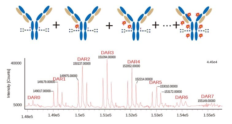
Figure 1. T-DM1 ADC DAR characterization results. T-DM1 is a mixture of ADCs with different DARs. With the LC-HRMS platform, the characterization of ADCs with different DARs can be realized.
How to Determine ADC DAR?
ADC analysis for DAR covers two key aspects: DAR distribution and average DAR. DAR distribution characterizes the proportion of ADC molecules with different DARs in the total ADC molecules. Average DAR is the ratio of the molar concentration of the total ADC molecules to that of the antibody molecule in the system. We have developed an LC-HRMS intact protein analysis to characterize the ADC DAR. Compared to hydrophobic interaction chromatography-ultraviolet (HIC-UV) and traditional DAR characterization methods, the LC-MS method can greatly reduce sample consumption and provide more accurate DAR results in ADC bioanalysis [1].
For ADCs in biological matrices, the standard ADC bioanalysis approach begins with immunoaffinity capture. Target ADCs are selectively enriched using the anti-human IgG, idiotype antibody, or antigen that can specifically bind to the analyte. Next, the LC-HRMS platform is used to conduct direct analysis on the purified ADC, and finally, deconvolution software is utilized for data processing (see Figure 2). Distinct peaks of ADC molecules with different absolute molecular weights and different DARs are obtained to complete the ADC DAR analysis.
Alternatively, purified ADC can be broken down to separate the light and heavy chains and then undergo an LC-HRMS assay. Once the deconvolution of the spectrogram is acquired, DARs of light and heavy chains can be obtained. The ADC DAR analysis can then be performed indirectly.
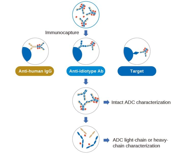
Figure 2. ADC DAR characterization process
This platform is used to evaluate the in vitro and in vivo stability of T-DM1. It provides a DAR analysis method with ultra-high sensitivity (LLOQ: 0.5-1 μg/mL), specificity and accuracy. The in vitro stability results are shown in Figure 3. The intact protein analysis results revealed the changes of DAR profile of T-DM1.
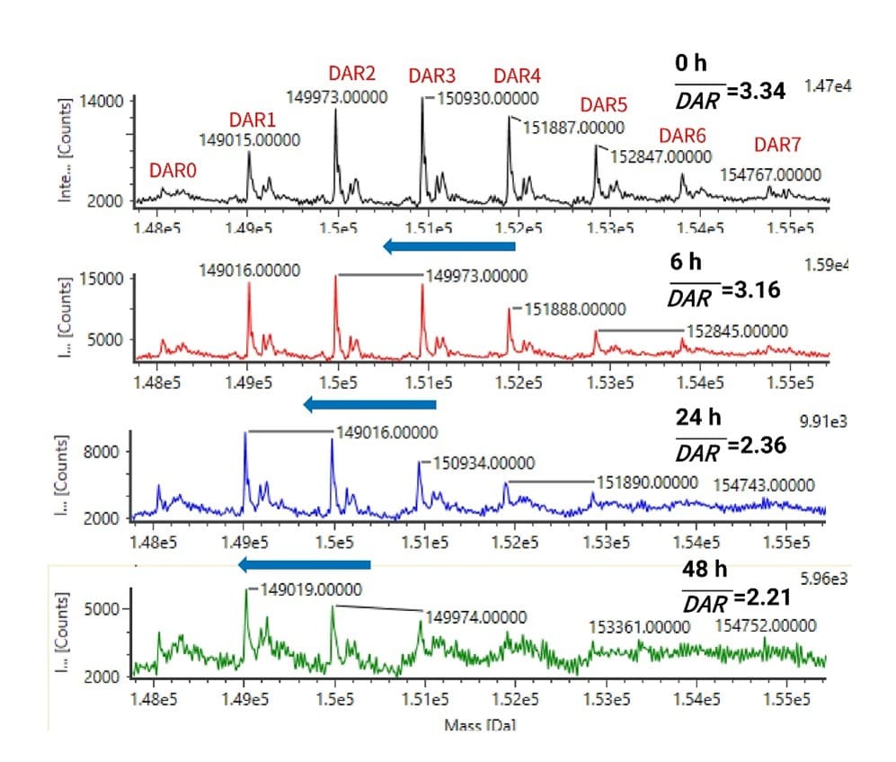
Figure 3. In vitro plasma stability of T-DM1. With the increase in incubation time, the content of high-DAR ADCs (DAR6 in the figure disappeared after 24 h) gradually decreased, and the average ADC DAR saw a corresponding decrease from 3.34 at T0 to 2.21 after 48 hours.
Quantitative Bioanalysis of ADC Drug Components in Serum/Plasma or Other Biological Matrices
Ligand Binding Assays (LBA)
The ligand binding assays (LBA) are the mainstream technique for measuring the antibody moiety of the ADCs, including total antibodies and conjugated antibodies. This method offers adequate sensitivity to analyze large molecules, alongside high throughput and robustness at a relatively low cost of implementation and instrumentation. And there is well-established guidance that can be referenced for the LBA method. Moreover, diverse LBA assay formats can be tailored to meet different assay requirements.
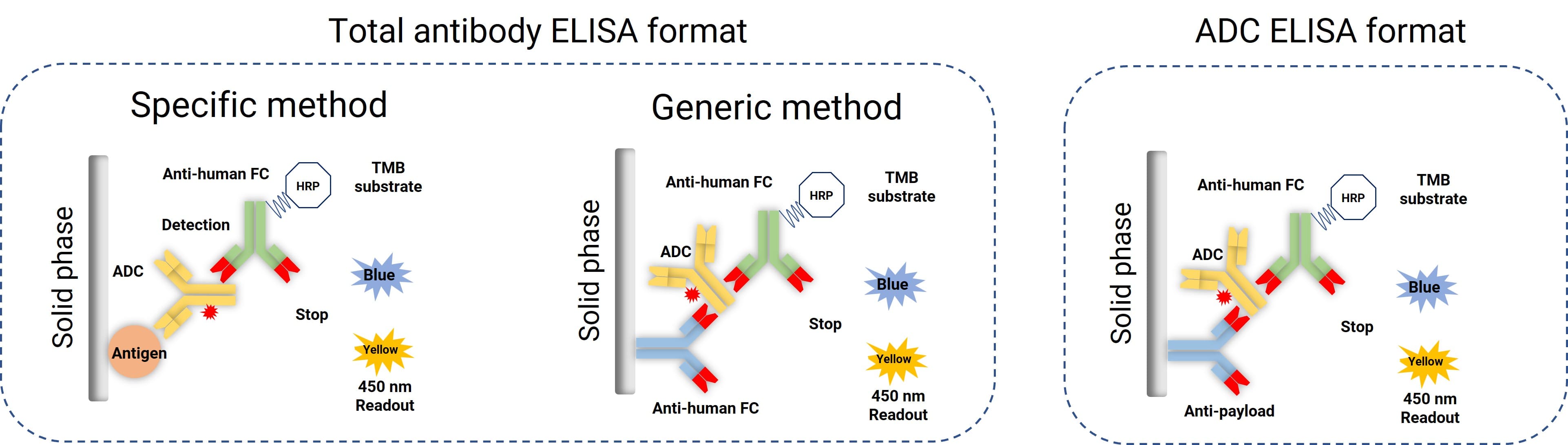
Figure 4. Common ELISA assay formats for total antibody and conjugated antibody bioanalysis
The ligand binding assay platform face several challenges specific to ADC bioanalysis. The conjugated antibody quantification generally cannot provide the ADC DAR information, nor can it detect ADC biotransformation. The specificity and sensitivity of LBA are highly dependent on the selection of critical reagents. The development of critical reagents for LBA can be costly and time-consuming, especially for anti-payload antibodies. Perhaps the most significant disadvantage in using LBA for ADC bioanalysis is their complete inability to capture information about the ADC's structure or possible biotransformation.
Liquid Chromatography-Mass Spectrometry (LC-MS)
In recent years, with the continuous development of mass spectrometry, LC-MS/MS and LC-HRMS have been increasingly used for macromolecular characterization and quantification [2-4]. Compared with the traditional ELISA, LC-MS offers a shorter development cycle and better standardization. In addition, LC-HRMS based on high-resolution mass spectrometry not only quantifies macromolecules but also determines the ADC DAR, thus facilitating the improvement of ADC analysis and biological characterization.
ADCs are often present in vivo as complex and dynamically changing mixtures caused by biotransformation, ADC DAR changes, or a combination of both. In this case, the existing PK is no longer clear with regards to the expression of “therapeutic concentration” over time, posing a unique challenge for quantitative ADC bioanalysis. As shown in Table 1, the components for analysis available to characterize ADC PK include total antibody (an antibody that is conjugated or unconjugated to the payload), conjugated antibody (ADC, antibody that is conjugated to at least one payload and is a prototype drug of ADC), unconjugated payload, conjugated payload and payload related species (effector formed after ADC cleavage or decomposition) [5].
The PK of different analytes reflects different content and significance, presenting the complete picture of ADC metabolism in vivo. Therefore, a comprehensive LC-MS platform is needed for the PK analysis of total antibodies, intact ADCs, unconjugated payload, and conjugated payload.
(1) Quantitative analysis of total antibody by LC-MS
For total antibody analysis of ADCs, sample preparation, and analytical workflows remain consistent with other antibody drugs. Usually, the process consists of three steps (Figure 5):
Immunoaffinity capture: the target total antibody is purified using antibodies or antigens that are specifically bound to the ADC antibody.
Proteolysis: antibody molecules are digested to generate the signature peptides.
LC-MS/MS detection: quantitative determination is performed via LC-MS/MS.
The determination of total antibody is quite consistent with the first step of DAR analysis. The only difference is that when the LC-MS/MS platform is used to determine the total antibody, the requirements for the specificity of the capture reagents are reduced, allowing for more cost-effective alternatives. Beyond anti-human IgG, idiotype antibodies, and antigens, Protein A/G is also a viable option. It offers broad universality at a lower price, albeit with relatively lower specificity
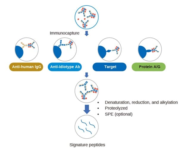
Figure 5. LC-MS/MS quantification process of total antibody in ADC bioanalysis
(2) Quantitative ADC analysis by LC-MS
It is important to begin this section by defining the target analyte of ADC quantitative analysis. There are generally two ways to define the PK target analyte of ADCs:
From the perspective of antibodies, it can be defined as antibody molecules bound to at least one drug.
From the perspective of drug load, it can be defined as the total concentration of the drug bound to the antibody.
Based on the two definitions above, we have developed two different ADC bioanalysis strategies. If an antibody molecule bound to more than one drug serves as the analyte, we can use a method similar to total antibody analysis. The difference is that the capture reagent used in the first step must be an anti-drug antibody. Therefore, the antibody molecules conjugated to drugs can be extracted and quantified using LC-MS/MS [2]. If the drug conjugated to the antibody is used as the analyte, and the linker can be cleaved through sample processing to release the drug, then the conjugated drug can be released after the purified total antibody is captured (Figure 6). Furthermore, the concentration of the conjugated drug can be determined to achieve the quantitative ADC bioanalysis.
In addition to the two situations noted above, through the steady development of site-specific conjugation [6], the “payload-linker-amino acid” generated by proteolysis has also been used as the analyte for ADC quantitative analysis in recent years. This makes it possible to achieve the quantitative analysis of conjugated payload. Theoretically, the average DAR of ADCs can be indirectly detected through the analysis of the quantitative results of conjugated payload and total antibody.
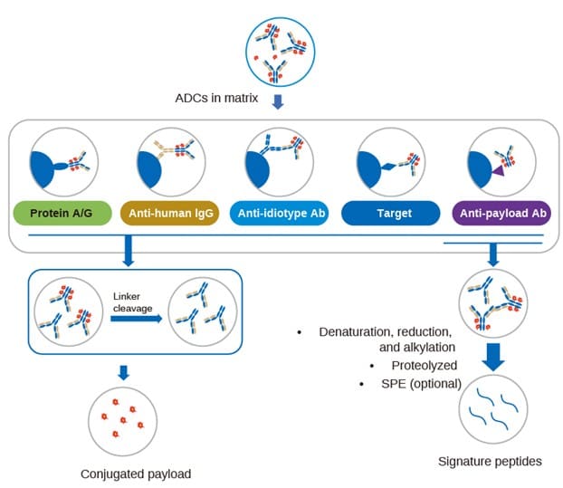
Figure 6. Quantitative strategy for ADC bioanalysis: 1) For non-cleavable linkers, the ADC can be captured by using an anti-payload antibody, and the antibody molecules conjugated to the drug can be quantified; 2) For cleavable linkers, the total concentration of the drug bound to the antibody can be quantified by releasing the conjugated payload.
(3) Quantitative ADC analysis of free payload by LC-MS
An important factor affecting ADC toxicity is the free payload. Since it may mediate unexpected off-target toxicity, monitoring free payload has become an indispensable part of ADC analysis. Due to its extremely low concentration in plasma, it presents a significant challenge regarding sensitivity. We have established quantitative bioanalytical methods using the highly sensitive AB 6500 and AB 7500 for the quantification of free payload. Figure 7 shows the LC-MS/MS chromatogram of MMAE at 20 pg/mL in the rat plasma sample. Based on the signal-to-noise ratio, LLOQ can be lower than 20 pg/mL linearity meets the method requirements. LC-MS/MS quantitative analysis of free payload is often applied in experiments to determine whether the free payload is released during ADC drug storage, linker stability in plasma, toxin stability in biological matrix, small molecule PK and tissue distribution, etc.
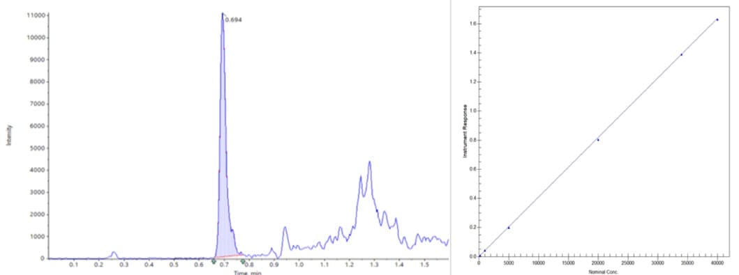
Figure 7. LC-MS/MS Chromatogram and Standard Curve of MMAE at 20 pg/mL in Rat Plasma Sample
Summary
Our ADC analysis team has established qualitative and semi-quantitative analysis methods for intact ADCs from plasma/serum or other biological matrices. These methods enable monitoring in vivo and in vitro changes in ADC DAR values and potential biotransformation. In addition, there is a well-established and comprehensive quantitative analysis protocol for various ADC components that can partially represent the PK characteristics, including total antibody, intact ADC, free payload, and conjugated payload (Figure 8). Notably, the intact ADC analysis method based on high-resolution mass spectrometry (HRMS)-based assay can even provide extremely extensive PK characteristic parameters of ADCs in just a single detection, including concentration and DAR. All these methods collectively constitute a complete set of ADC bioanalysis strategies for ADCs.
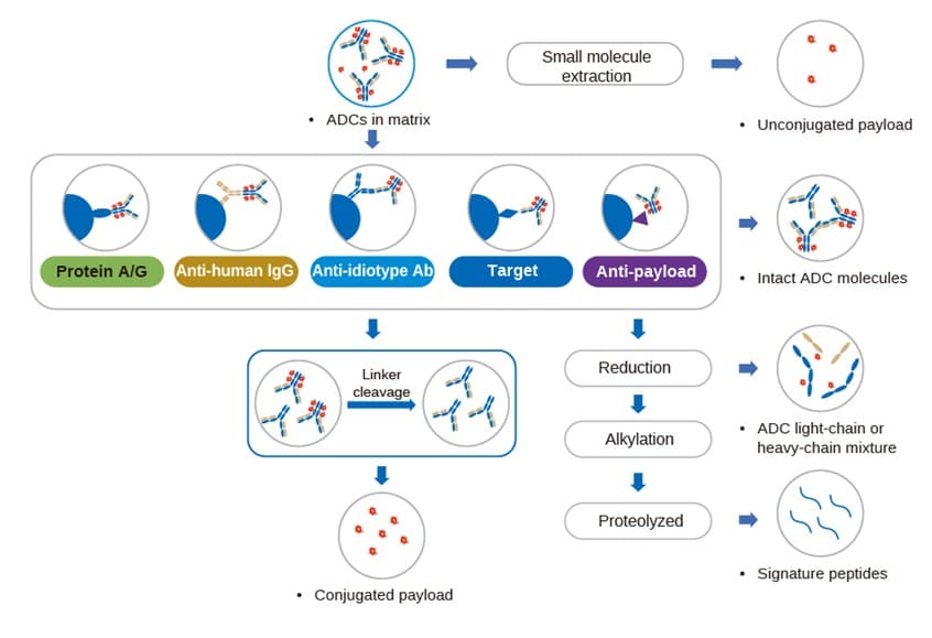
Figure 8. Summary diagram of LC-MS assay strategy for ADC-related components [7]
Author: Maotian Zhou, Huan Yan, Li Qu, Zhiyu Li, Lili Xing
Talk to a WuXi AppTec expert today to get the support you need to achieve your drug development goals.
Committed to accelerating drug discovery and development, we offer a full range of discovery screening, preclinical development, clinical drug metabolism, and pharmacokinetic (DMPK) platforms and services. With research facilities in the United States (New Jersey) and China (Shanghai, Suzhou, Nanjing, and Nantong), 1,000+ scientists, and over fifteen years of experience in Investigational New Drug (IND) application, our DMPK team at WuXi AppTec are serving 1,600+ global clients, and have successfully supported 1,800+ IND applications.
Reference
[1] Källsten, M., Hartmann, R., Artemenko, K., et al. (2018). Qualitative analysis of antibody–drug conjugates (ADCs): an experimental comparison of analytical techniques of cysteine-linked ADCs. Analyst, 143(22), 5487-5496.
[2] Huang, Y., Mou, S., Wang, Y., et al. (2021). Characterization of Antibody–Drug Conjugate Pharmacokinetics and in Vivo Biotransformation Using Quantitative Intact LC-HRMS and Surrogate Analyte LC-MRM. Analytical Chemistry, 93(15), 6135-6144.
[3] Jin, W., Burton, L., & Moore, I. (2018). LC–HRMS quantitation of intact antibody drug conjugate trastuzumab emtansine from rat plasma. Bioanalysis, 10(11), 851-862.
[4] Kellie, J. F., Pannullo, K. E., Li, Y., et al. (2020). Antibody subunit LC-MS analysis for pharmacokinetic and biotransformation determination from in-life studies for complex biotherapeutics. Analytical Chemistry, 92(12), 8268-8277.
[5] Zhu, X., Huo, S., Xue, C., et al. (2020). Current LC-MS-based strategies for characterization and quantification of antibody-drug conjugates. Journal of pharmaceutical analysis, 10(3), 209-220.
[6] Lee, B. I., Park, M. H., Byeon, J. J., et al. (2020). Quantification of an Antibody-Conjugated Drug in Fat Plasma by an Affinity Capture LC-MS/MS Method for a Novel Prenyl Transferase-Mediated Site-Specific Antibody–Drug Conjugate. Molecules, 25(7), 1515.
[7] Liang Shen, editor. Drug Metabolism and Pharmacokinetics: Frontiers, Strategies, and Applications. Wiley. 2025. http://doi.org/10.1002/9781394300150.


Related Services and Platforms


-
 DMPK BioanalysisLearn More
-
Novel Drug Modalities DMPK Enabling PlatformsLearn More
-
Novel Drug Modalities BioanalysisLearn More
-
Small Molecules BioanalysisLearn More
-
Bioanalytical Instrument PlatformLearn More
-
PROTAC DMPK ServicesLearn More
-
ADC DMPK ServicesLearn More
-
Antibody-Oligonucleotide Conjugate (AOC) DMPK ServicesLearn More
-
Oligo DMPK ServicesLearn More
-
PDC DMPK ServicesLearn More
-
Peptide DMPK ServicesLearn More
-
mRNA DMPK ServicesLearn More
-
Covalent Drugs DMPK ServicesLearn More
Stay Connected
Keep up with the latest news and insights.

