



Evaluating the In Vitro Stability of Antibody-Drug Conjugates in Blood Matrixes
-

Articles
-

Sep 17, 2026
Why Does In Vitro Stability Matter for Antibody–Drug Conjugates?
Antibody-drug conjugates (ADCs) typically consist of a monoclonal antibody (mAb) covalently linked to a cytotoxic payload via a chemical ADC linker [1]. The antibody helps to deliver payload to the disease site via targeting a specific antigen expressed on the surface of the cancer cells. Therefore, ADCs can decrease the off-target toxicity to normal tissues. It combines both the advantages of highly specific targeting ability and highly potent activity to achieve precise and efficient elimination of cancer cells, which has become one of the hotspots for the research and development of anticancer drugs [2].
The ADC stability in circulation is critical for maximum efficacy and minimal toxicity. An intact ADC reaching the intended target can deliver the highest possible drug load to the tumor and reduce off-target toxicity caused by the premature release of the payload in the blood [2]. Several factors may impact the ADC stability, including linker [3], conjugation strategy [4], physicochemical properties of the payload [5], drug-to-antibody ratio (DAR) [6], etc. It is a relatively simple and direct method to assess the in vitro ADC stability of using blood matrixes. The National Medical Products Administration (NMPA) issued The Technical Guideline for Non-clinical Studies of Antibody-Drug Conjugates in September 2023 [7], which stated that “Before starting the clinical trials, the in vitro stability of ADCs should be investigated in plasma and/or blood matrixes in humans and animals (pharmacology and/or toxicology species). This study is required to select appropriate analytes based on the release characteristics of ADCs, such as ADC remaining and/or the generation of free payload.”
This article introduces various in vitro incubation systems and stability results, and summarizes the in vitro plasma/serum stability studies of all marketed ADCs. It is intended as a reference for in vitro ADC stability assessment in ADC development and pharmacokinetic investigations.
How to Evaluate In Vitro ADC Stability in Plasma
Plasma is the most commonly used blood matrix for evaluating the in vitro stability of ADCs. It is well known that plasma is obtained by centrifugation of blood containing anticoagulant, in which the blood cells are removed. Moreover, plasma contains many kinds of hydrolytic enzymes [8].
Plasma has been widely used in various preclinical and clinical studies due to its advantages, such as simple and quick preparation, high yield, and convenience of frozen storage and transportation. In fact, the anticoagulant selection requires attention in the plasma preparation. Ethylenediaminetetraacetic acid (EDTA), a commonly used anticoagulant, is an inhibitor of proteases and nucleases. Consequently, plasma prepared with EDTA may mislead the stability results of ADCs that are sensitive to proteases, and sodium heparin is therefore required as the anticoagulant. To mimic in vivo conditions, the candidate molecule is typically incubated at 37°C. The ADC remaining and/or the generation of free payload are determined at different time points. The incubation time generally varies from 4 to 21 days, and sterility should be maintained during the incubation. The selected species should include human and intended pharmacology and toxicology species, and the incubated ADC concentrations are recommended to be close to the observed or predicted in vivo Cmax [9]. In addition, the negative control would typically include appropriate buffers (e.g., phosphate buffer saline (PBS)) with protein (e.g., bovine serum albumin (BSA)) to determine the ADC stability under buffer systems [10].
So far, literature data indicate that in vitro plasma stability of many ADCs translates well with in vivo results. In the study of Enhertu (DS8201), 1.2% to 3.9% of DXd was detected in mouse, rat, monkey, and human plasma on day 21 (Figure 1), indicating that Enhertu is stable in plasma. For another marketed ADC of Polivy, the concentrations of total antibody significantly decreased within 24 hours, while the decrease was much slower after 24 hours (Figure 2 A); At 96 hours, the conjugated payload decreased to around 60% (Figure 2 B) [12].
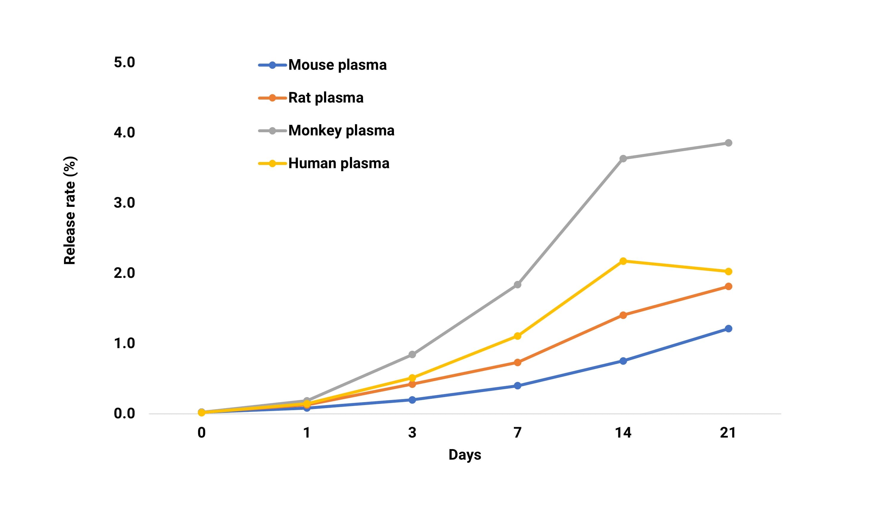
Figure 1. In vitro stability of DS-8201 in plasma. Data from reference [11]
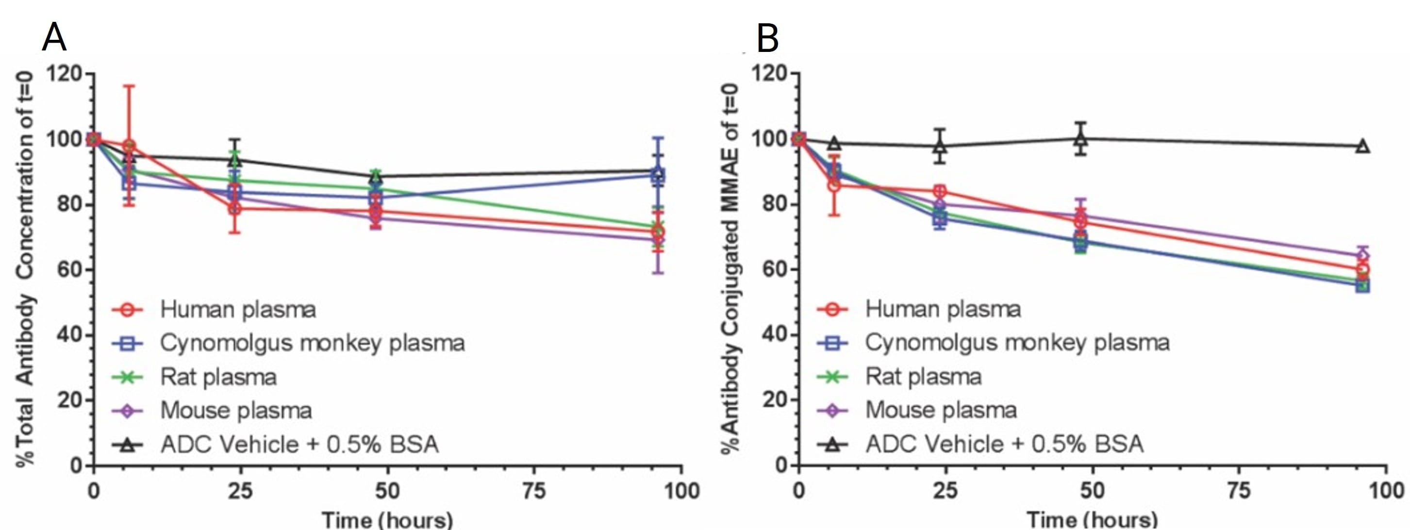
Figure 2. Total antibody (A) and antibody conjugated MMAE (B) levels of Polivy following 96 hours of incubation in human, cynomolgus monkey, rat, and mouse plasma at 37°C [12]
In the plasma stability study of maleimide-conjugated ADC of Tras-mcVC-auristatin, Han et al. [13] observed a discrepancy between the measured free payload concentrations and loss of DAR (Figure 3). The changes in DAR values suggested that the ADC had lost 30−50% of its payload, while free payload concentrations measured in plasma showed that very little loss of payload occurred. Further studies suggested that the deconjugated maleimide linker payload is transferred to serum albumin to form adducts, causing the decrease in DAR. However, this part of the payload cannot be measured in the form of free payload, which also indicates that there is a certain risk in assessing the ADC stability by only detecting free payload generation.
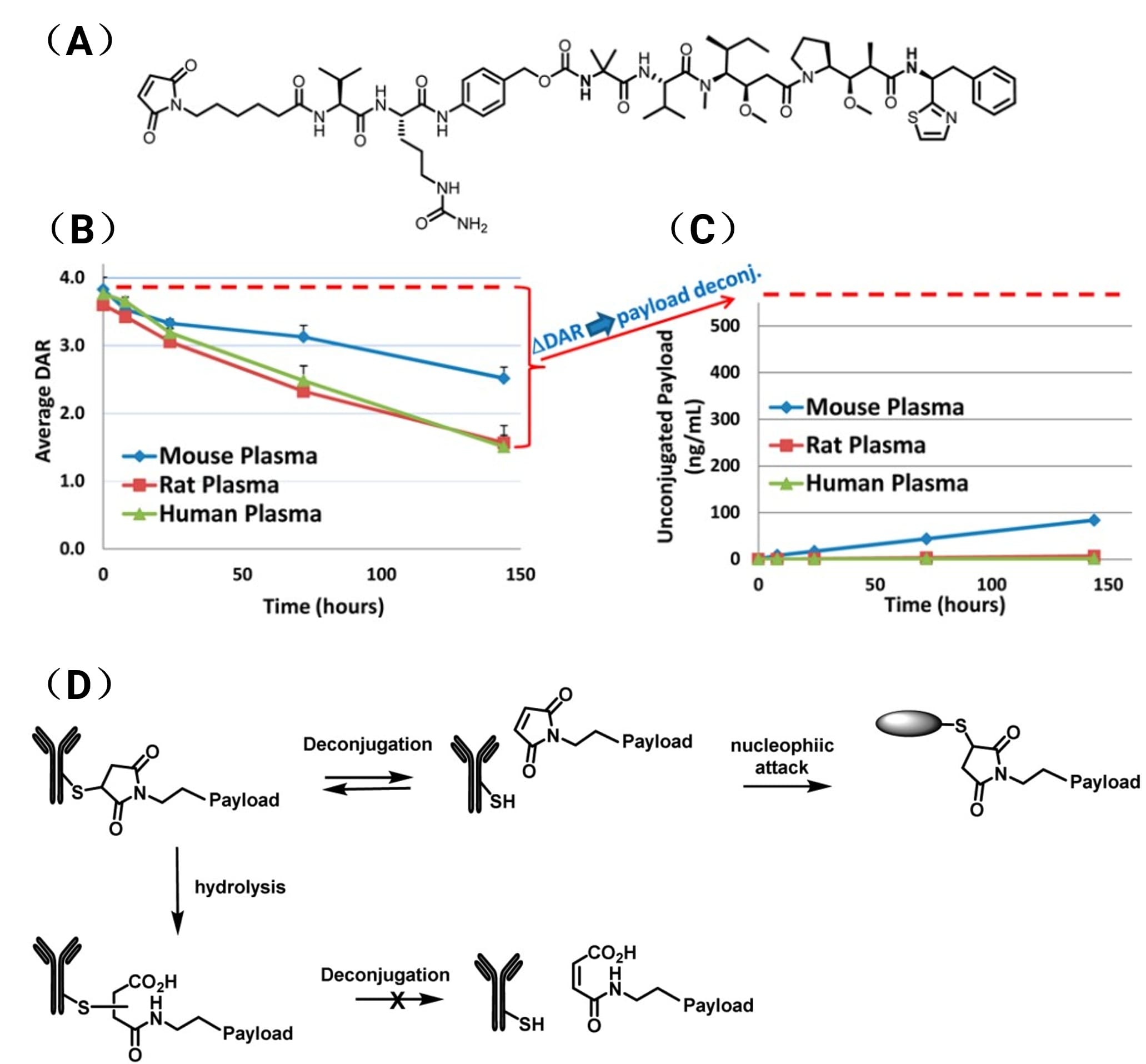
Figure 3. (A) Chemical structure of linker-payload of Tras-mcVC-Auristatin ADC; (B) Average DAR profiles of the Tras-mcVC-Auristatin ADC over the time-course of the in vitro plasma incubation; and (C) Unconjugated/free payload levels of Tras-mcVC-Auristatin over the time-course of the in vitro plasma incubation; (D) Scheme of the maleimide exchange from the ADC [13]
It has been reported that the frozen plasma will reduce the activity of enzymes involved in ADC linker-payload modification [14]. Therefore, caution is needed for studies using frozen plasma. Inconsistency in stability in vitro and in vivo has also been reported, for example, Dorywalska et al. found that the VC-PABC linker appears to be cleaved more efficiently in vivo compared with in vitro plasma stability assay in mice. The researchers rationalized that this could be due to a continuous enzyme source in vivo or the contribution of intracellular degradation [15].
How to Evaluate In Vitro ADC Stability in Serum
Serum is the clear liquid that separates from blood when blood is allowed to clot completely and then centrifuged. Therefore, the serum contains no fibrinogen and no anticoagulants. Compared with plasma, using serum can circumvent the potential risks associated with the inhibition of enzyme activity by anticoagulants, as well as the effect on protein binding and stability in serum [8]. Serum is also commonly used for in vitro ADC stability studies. The study design of incubation conditions and detection is similar to plasma stability.
Aidixi was incubated in human, rat, and mouse serum for 24 hours at 4°C and 37°C, separately, and the release of MMAE was detected. The results showed that the release of MMAE in mouse serum was 4.3% (at 37°C), and less than 1% under other conditions. Aidixi was most stable in human serum, followed by rats and mice, according to their MMAE release, indicating species differences. Overall, Aidixi showed good serum stability [16]. In the study of Trodelvy, free SN-38 concentrations were measured after 120 hours of incubation in human and monkey serum. The results showed that the half-life of SN-38 release in serum in vitro was approximately 1 day [17].
How Does Whole Blood Perform as a Matrix for ADC Stability?
Whole blood is considered to better mimic the in vivo circulatory environment, which includes blood cells and plasma. Whole blood has been included as one of the recommended matrixes for the in vitro ADC stability study in the NMPA guidelines. Different from plasma and serum, whole blood cannot be frozen at -80°C and is inconvenient for storage and transportation. The stability study requires a fresh matrix to be performed. Up to now, there are only a few published reports on the in vitro ADC stability using whole blood as the matrix.
Aimee et al. used a variety of THIOMABTM antibody-drug conjugates (TDCs) to evaluate the ADC stability both in whole blood and plasma, and to correlate in vitro data to in vivo data [14]. Results of whole blood showed an improved correlation to in vivo stability (R2 = 0.87, coefficient of determination) compared with those of unfrozen or frozen mouse plasma (R2 = 0.34, 0.01, respectively). Further studies showed that the whole blood assay can also predict in vivo stability in other preclinical species (e.g., rat and cynomolgus monkey) as well as in humans. Based on our research experience, red blood cells may be broken if blood is incubated in vitro for over 24 hours. For the ADCs that require more than 24 hours of incubation, the applicability of whole blood needs to be further evaluated.
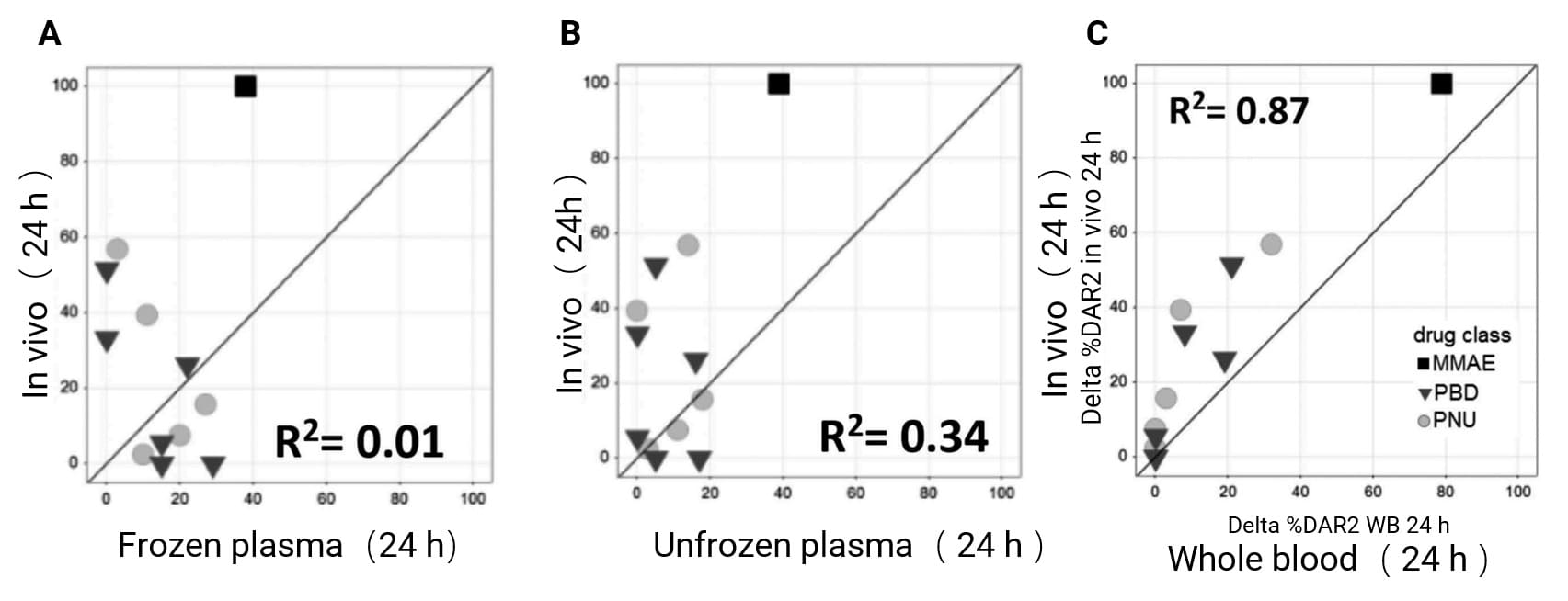
Figure 4. Correlation of in vivo with in vitro stability for 13 TDCs in frozen mouse plasma (A), unfrozen mouse plasma (B), and mouse whole blood at 24 hours [14]
Note: Axes represent percent loss of drug relative to 0 hour
Analyte Selection for ADC Bioanalysis In Vitro Stability Studies
There are multiple options for the analytes in the in vitro ADC stability study. Various analytes and their characteristics are summarized as follows.
Free payload: The in vitro ADC stability is evaluated by detecting the free payload released at different time points. However, it is difficult to determine payload release when the free payload is unstable in blood matrixes or easily covalently bound to proteins. In addition, the sensitivity of detection is required to be high enough, and quantitative ADC bioanalysis is recommended.
ADC: ADC concentration is generally measured by ligand binding assays (LBA). Since conjugated antibodies with one or more payloads will be treated as the ADC in LBA, measuring the ADC concentrations by LBA cannot sensitively evaluate the payload release. Besides, anti-payload antibodies, which are necessary for ADC detection by the LBA method, are usually not available at an early stage for ADCs conjugated with new payloads.
DAR value: ADC stability is assessed by detecting changes in DAR over time. The average DAR values can sensitively evaluate ADC stability. Payload release from ADC will cause the DAR value changes, no matter what components are being released or whether the payload is stable or not, DAR detection can effectively avoid those issues and it is more recommended to evaluate ADC stability.
Total antibody: This method is mainly used to evaluate the stability of antibodies. By comparing the differences between the conjugated antibody and total antibody, we can further analyze whether the instability of ADC is caused by payload release or the instability of the antibody itself.
Conjugated payload: That is, detecting the concentration of payload conjugated to ADC. It requires purification of ADC using immunocapture first, then followed by hydrolyzing the payload from ADC, which is applied to cleavable ADCs but not non-cleavable ADCs. Hydrolyzing the payload effectively from ADC is the most challenging part of sample pretreatment and requires case-by-case optimization of hydrolysis conditions.
Summary of In Vitro Stability Studies of Marketed ADCs
22 ADCs have been approved worldwide until 2026, and in vitro stability studies have been conducted in many of them. Here we summarize the information in Table 1. For most marketed ADCs, plasma was selected to evaluate the in vitro stability, and a buffer group containing BSA was also included. Compared with conventional small molecules, the incubation time in ADC plasma stability study is longer, varying from 1 to 21 days. By detecting the concentration of free payload and/or total antibody, the payload release or the instability of the antibody part is evaluated. Among all the approved ADCs, serum was only used to evaluate in vitro stability for Trodelvy and Aidixi.
Table 1. Summary of In Vitro Stability Study Design for Marketed ADCs [18-21]
| Drug | Matrix | Species | Incubation Time | Incubation Drug | Analyte |
1 | Mylotarg | NA |
|
|
|
|
2 | Adcetris | Plasma | R, C, H | 21 days | ADC | MMAE |
3 | Kadcyla | Plasma | R, C, H | 4 days | Tmab-MCC- [3H] DM1 | Radioactivity, ADC/Total Antibodies in Acetonitrile-Treated Supernatant and Precipitate |
4 | Besponsa | Plasma | R, C, H | 4 days | [3H] Inotuzumab Ozogamicin [3H] N-Ac-γ-calicheamicin DMH | Calicheamicin |
5 | Lumoxiti | NA |
|
|
|
|
6 | Polivy | Plasma | C, M, R, H | 4 days | ADC | Total Antibodies, MMAE |
7 | Padcev | NA |
|
|
|
|
8 | Enhertu | Plasma | C, M, R, H | 21 days | ADC | DXd |
9 | Trodelvy | Serum | C, H |
| ADC | SN-38 |
10 | Blenrep | Plasma | C, R, H | 4 days | ADC | McMMAF |
11 | Akalux | NA |
|
|
|
|
12 | Zynlonta | Plasma | C, H | 7 days | ADC |
|
13 | Tivdak | Plasma, | M, C, H | 14 days | ADC | MMAE |
14 | Aidixi | Serum | H, R, M | 1 day | ADC | MMAE |
15 | Elahere | NA |
|
|
|
|
16 | Sacituzumab tirumotecan | Plasma | C, H | 6 days | ADC | KL610023 |
17 | Datroway | Plasma | C, M, R, H | NA | ADC | Dxd |
18 | Emrelis | NA |
|
|
|
|
19 | Trastuzumab rezetecan | Plasma | C, M, R, H | 21 days | ADC | SHR9265 |
20 | Trastuzumab botidotin | NA |
|
|
|
|
21 | Becotatug vedotin | NA |
|
|
|
|
22 | Izalontamab brengitecan | NA |
|
|
|
|
Note: C: monkey; M: mouse; R: rat; H: human
Summary
In ADC drug development, except for the selection of antibody and payload, it is also important to incorporate an appropriate linker to combine both parts and comprehensively consider the effects of various factors on the stability, toxicity, pharmacokinetics, and pharmacodynamics of ADCs. The in vitro stability study in blood matrixes can help to assess ADC stability in the circulatory system, off-target risk, and species differences.
Authors: Xinmeng Shi, Xiangling Wang, Jing Jin
Talk to a WuXi AppTec expert today to get the support you need to achieve your drug development goals.
Committed to accelerating drug discovery and development, we offer a full range of discovery screening, preclinical development, clinical drug metabolism, and pharmacokinetic (DMPK) platforms and services. With research facilities in the United States (New Jersey) and China (Shanghai, Suzhou, Nanjing, and Nantong), 1,300+ scientists, and over fifteen years of experience in Investigational New Drug (IND) application, our DMPK team at WuXi AppTec are serving 1,700+ global clients, and have successfully supported 2,100+ IND applications.
Reference
[1] Samantasinghar A, Sunildutt NP, Ahmed F, et al. A comprehensive review of key factors affecting the efficacy of Antibody-Drug Conjugates. Biomed Pharmacother. 2023 May; 161:114408.
[2] Tsuchikama K, An Z. Antibody-drug conjugates: recent advances in conjugation and linker chemistries. Protein Cell. 2018 Jan; 9 (1): 33-46.
[3] Su Z, Xiao D, Xie F, et al. Antibody-drug conjugates: Recent advances in linker chemistry. Acta Pharm Sin B. 2021 Dec; 11 (12): 3889-3907.
[4] Zhou Q. Site-Specific Antibody Conjugation for ADC and Beyond. Biomedicines. 2017 Nov 9; 5 (4): 64.
[5] Shen BQ, Xu K, Liu L, et al. Conjugation site modulates the in vivo stability and therapeutic activity of antibody-drug conjugates. Nat Biotechnol. 2012 Jan 22; 30 (2): 184-9.
[6] Sun X, Ponte JF, Yoder NC, et al. Effects of Drug-Antibody Ratio on Pharmacokinetics, Biodistribution, Efficacy, and Tolerability of Antibody-Maytansinoid Conjugates. Bioconjug Chem. 2017 May 17; 28 (5): 1371-1381.
[7] NMPA Guidelines: Technical Guidelines for Nonclinical Studies of Antibody-Conjugated Drugs, 25 September 2023
[8] Uges DR. Plasma or serum in therapeutic drug monitoring and clinical toxicology. Pharm Weekbl Sci. 1988 Oct 14; 10 (5): 185-8.
[9] Kraynov E, Kamath AV, Walles M, et al. Current Approaches for Absorption, Distribution, Metabolism, and Excretion Characterization of Antibody-Drug Conjugates: An Industry White Paper. Drug Metab Dispos. 2016 May; 44 (5): 617-23.
[10] Fisher JE Jr. Considerations for the Nonclinical Safety Evaluation of Antibody-Drug Conjugates. Antibodies (Basel). 2021 Apr 19; 10 (2): 15.
[11] ENHERTU (fam-trastuzumab deruxtecan-nxki). NDA/BLA Multi-disciplinary Review and Evaluation (BLA 761139).2020.
[12] Li D, Lee D, Dere RC, et al. Evaluation and use of an anti-cynomolgus monkey CD79b surrogate antibody-drug conjugate to enable clinical development of polatuzumab vedotin. Br J Pharmacol. 2019 Oct; 176 (19): 3805-3818. And its supplement.
[13] Wei C, Zhang G, Clark T, et al. Where Did the Linker-Payload Go? A Quantitative Investigation on the Destination of the Released Linker-Payload from an Antibody-Drug Conjugate with a Maleimide Linker in Plasma. Anal Chem. 2016 May 3; 88 (9): 4979-86.
[14] Fourie-O 'Donohue A, Chu PY, Dela Cruz Chuh J, et al. Improved translation of stability for conjugated antibodies using an in vitro whole blood assay. MAbs. 2020 Jan-Dec; 12 (1): 1715705.
[15] Dorywalska M, Strop P, Melton-Witt JA, et al. Site-Dependent Degradation of a Non-Cleavable Auristatin-Based Linker-Payload in Rodent Plasma and Its Effect on ADC Efficacy. PLoS One. 2015 Jul 10; 10 (7): e0132282.
[16] Li L, Xu MZ, Wang L, et al. Conjugating MMAE to a novel anti-HER2 antibody for selective targeted delivery. Eur Rev Med Pharmacol Sci. 2020 Dec; 24 (24): 12929-12937.
[17] Goldenberg DM, Cardillo TM, Govindan SV, et al. Trop-2 is a novel target for solid cancer therapy with sacituzumab govitecan (IMMU-132), an antibody-drug conjugate (ADC). Oncotarget. 2015 Sep 8; 6 (26): 22496-512.
[18] Liang Shen, editor. Drug Metabolism and Pharmacokinetics: Frontiers, Strategies, and Applications. Wiley. 2025. http://doi.org/10.1002/9781394300150.
[19] Cheng Y, Yuan X, Tian Q, et al. Corrigendum: Preclinical profiles of SKB264, a novel anti-TROP2 antibody conjugated to topoisomerase inhibitor, demonstrated promising antitumor efficacy compared to IMMU-132. Front Oncol. 2023;13:1334938.
[20] Swiss Public Assessment Report-DATROWAY.
[21] Zhang T, Xu J, Yin J, et al. SHR-A1811, a novel anti-HER2 antibody-drug conjugate with optimal drug-to-antibody ratio, efficient tumor killing potency, and favorable safety profiles. PLoS One. 2025;20(6):e0326691. pii: PONE-D-24-26156.


Related Services and Platforms


-
 In Vitro ADME ServicesLearn More
-
Novel Drug Modalities DMPK Enabling PlatformsLearn More
-
Physicochemical Property StudyLearn More
-
Permeability and Transporter StudyLearn More
-
Drug Distribution and Protein Binding StudiesLearn More
-
Metabolic Stability StudyLearn More
-
Drug Interactions StudyLearn More
-
PROTAC DMPK ServicesLearn More
-
ADC DMPK ServicesLearn More
-
Antibody-Oligonucleotide Conjugate (AOC) DMPK ServicesLearn More
-
Oligo DMPK ServicesLearn More
-
PDC DMPK ServicesLearn More
-
Peptide DMPK ServicesLearn More
-
mRNA DMPK ServicesLearn More
-
Covalent Drugs DMPK ServicesLearn More


Stay Connected
Keep up with the latest news and insights.

