



Magnetic Solid-Phase Extraction (mSPE) for Oligonucleotide Bioanalysis: Principles, Applications, and Strategies
-

Articles
-

Nov 12, 2026
In pharmacokinetic and toxicokinetic studies of oligonucleotide therapeutics, liquid chromatography–tandem mass spectrometry (LC-MS/MS) has become a mainstream bioanalytical platform because of its high specificity and sensitivity [1–3]. However, highly sensitive detection depends on efficient sample preparation, the primary objectives of which are to remove interfering substances from biological matrices, such as plasma, urine, and tissues, and to enrich and concentrate the target oligonucleotide for accurate bioanalysis.
Oligonucleotide therapeutics are highly water-soluble, carry substantial negative charge, and readily bind nonspecifically to proteins. These physicochemical properties present distinctive sample-preparation challenges. Conventional protein precipitation (PPT) does not provide effective extraction. Although liquid–liquid extraction (LLE) is straightforward to perform, endogenous interferents are readily co-extracted, resulting in pronounced matrix effects, particularly when processing tissue homogenates and excretory matrices. Solid-phase extraction (SPE) provides better sample cleanup, but the procedure is complicate and time-consuming.
To overcome these limitations, magnetic solid-phase extraction (mSPE) offers a new approach. By combining the advantages of magnetic carriers and solid-phase extraction, this technique provides effective sample cleanup while facilitating automation, demonstrating substantial application potential. Beginning with the principles of magnetic extraction, this article systematically describes three core interaction mechanisms used to extract oligonucleotide therapeutics—hybridization capture (complementary base pairing), ion exchange, and hydrophobic interactions—and their applicable settings. It also compares the performance of different sample-preparation approaches through a practical case study, providing a reference for selecting bioanalytical methods for oligonucleotide therapeutics.
Principles and Technical Advantages of Magnetic Solid-Phase Extraction
Magnetic solid-phase extraction (mSPE) is a novel dispersive solid-phase extraction technique based on liquid–solid chromatography principles. It uses magnetic or magnetizable materials as sorbents and enables rapid enrichment of target analytes with an external magnetic field during the bioanalysis of oligonucleotides. The key material—the magnetic sorbent, or magnetic bead—consists of an iron oxide magnetic core and a shell coated with specific functional groups. Differences in functional-group structure determine the mechanism and strength of interactions with the analyte, thereby affecting extraction or elution efficiency. Figure 1 illustrates the interactions between oligonucleotide therapeutics and magnetic beads bearing different functional groups.
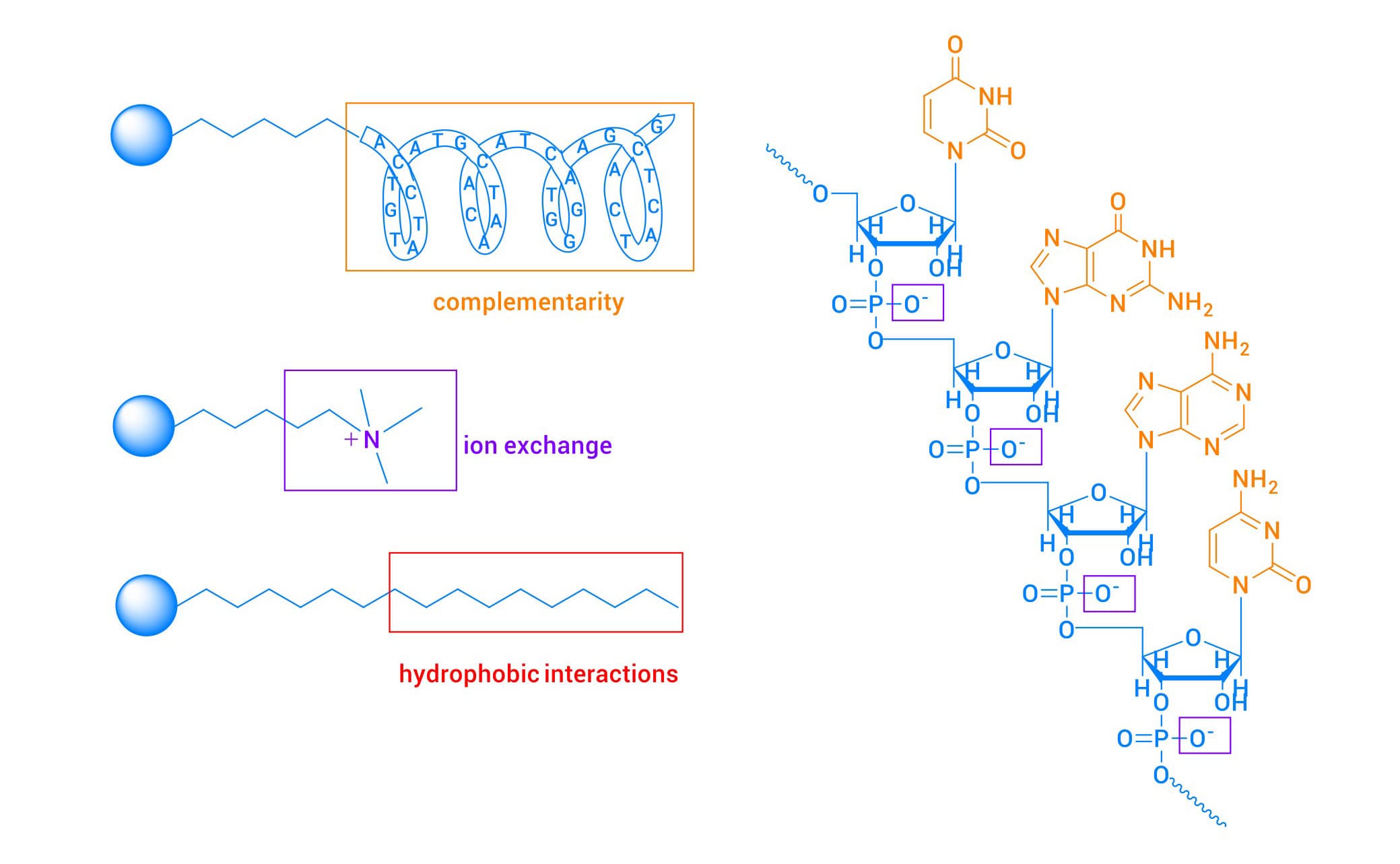
Figure 1. Schematic illustration of interactions between oligonucleotides and magnetic beads bearing different functional groups
The basic principle of mSPE is shown in Figure 2 [4]. A magnetic sorbent is added to the sample solution or suspension, and the target analytes adsorb onto the surface of the dispersed sorbent through specific interactions. Under an external magnetic field, the target analytes migrate together with the sorbent. Impurities are removed with a wash solution, after which the target analytes are eluted with an appropriate elution solvent, achieving sample enrichment and purification.
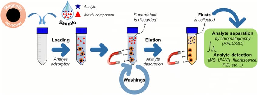
Figure 2. Schematic illustration of the principle of magnetic solid-phase extraction [4]
Compared with conventional LLE and SPE, mSPE offers the following notable advantages for oligonucleotide bioanalysis:
High mass-transfer efficiency: Magnetic beads have a large specific surface area and rapid mass transfer, allowing target compounds to be adsorbed from large sample volumes within a short time.
Operational convenience: Rapid separation and collection can be achieved by applying an external magnetic field, avoiding cumbersome filtration or centrifugation steps and facilitating automation.
High sample cleanliness: The extracts offer clear advantages in cleanliness and specificity, effectively reducing matrix effects.
The technique is now widely used in biomedicine, food safety, and environmental monitoring.
Core Mechanisms of mSPE: Hybridization Capture, Ion Exchange, and Hydrophobic Interactions
Based on the physicochemical properties of oligonucleotides, magnetic extraction techniques can be classified into three categories according to their interaction mechanisms: hybridization capture (complementary base pairing), ion exchange, and hydrophobic interactions. These three extraction modes have different performance strengths. An appropriate approach can be selected according to the analytical objectives to achieve flexible and efficient sample preparation.
Complementary Base Pairing (Hybridization Capture)
Principle
Complementary base pairing, also known as nucleic acid hybridization, is the process in which two single-stranded nucleic acids with a certain degree of sequence homology reanneal under appropriate conditions to form a duplex through complementary base pairing.
The workflow for nucleic acid hybridization capture using a PNA probe is shown in Figure 3 [5]:
Heat-denature the double-stranded siRNA in the sample.
Add a biotin-labeled capture probe. At an appropriate ionic strength and annealing temperature, the probe specifically hybridizes to the target sequence through complementary base pairing.
Add streptavidin-coated magnetic beads. The specific affinity between biotin and streptavidin produces magnetic bead–probe–target nucleic acid complexes.
Retain the beads by magnetic separation and wash repeatedly with wash buffer to remove unbound non-target nucleic acids and impurities.
Use elution buffer and elevated temperature to dissociate the capture probe from the target nucleic acid and recover the enriched target nucleic acid.
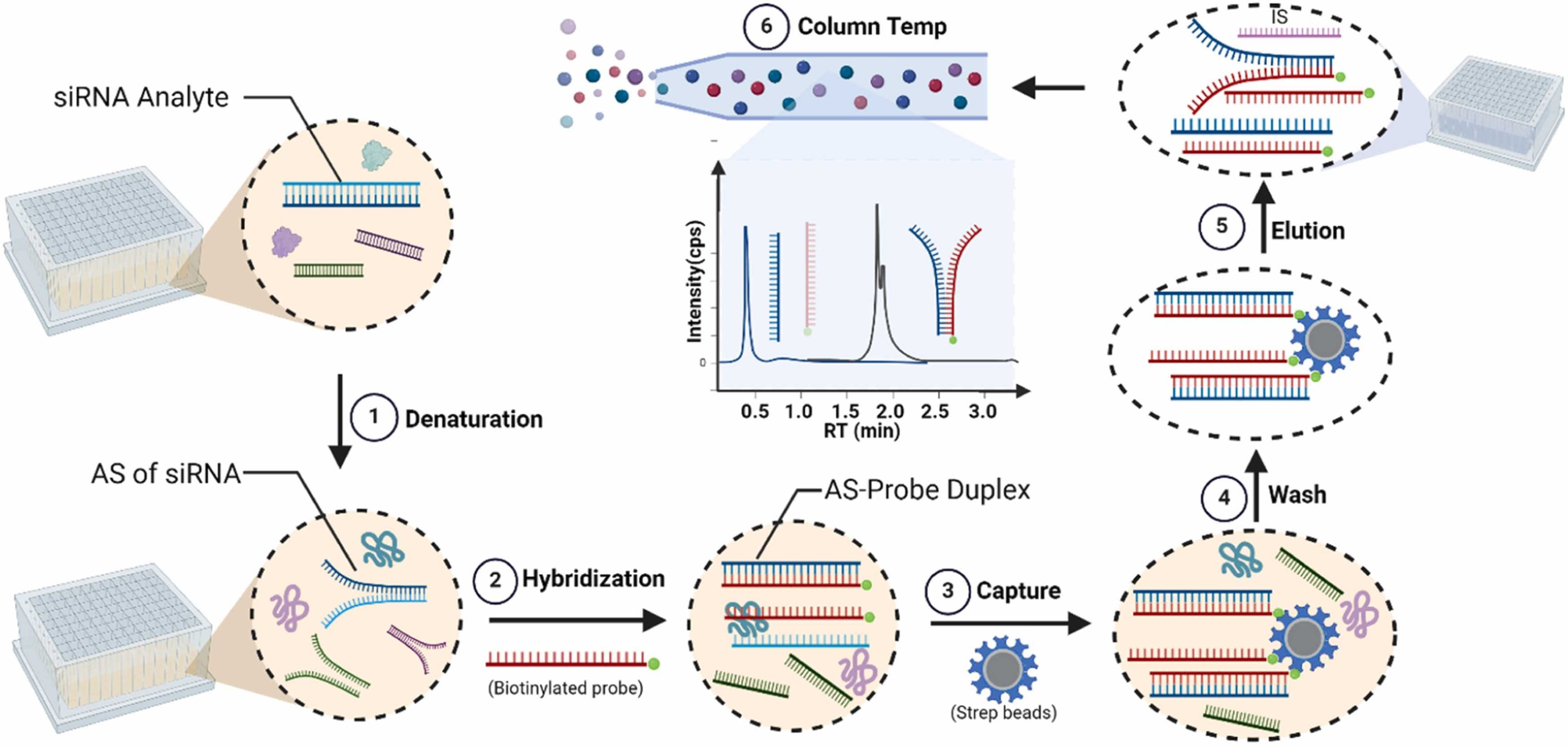
Figure 3. Schematic workflow for PNA-probe-based nucleic acid hybridization capture [5]
Technical Features
The principal advantages of this method are:
High specificity: Stringent complementary base pairing enables precise recognition of the target sequence, allowing homologous sequences to be distinguished and cross-reactivity to be avoided in DMPK studies.
High sensitivity: Trace amounts of DNA/RNA can be detected, making the method suitable for low-abundance samples.
High operational safety: Reactions are performed in an aqueous system under mild conditions, using a standardized workflow.
Main Challenges
Stringent experimental requirements: Probe design, reaction temperature, reagent quality, and salt concentration can substantially affect reproducibility in the bioanalysis of oligonucleotides.
Longer processing time: Multiple steps, including denaturation, hybridization, washing, and elution, are required, limiting throughput.
Key Influencing Factors
The following parameters require particular attention during method development:
Probe design: Use an appropriate probe length and base composition; avoid dimer or hairpin formation; and ensure highly specific matching to the target sequence.
Hybridization temperature: Control the temperature closely, generally with reference to the melting temperature (Tm). Excessively high temperatures reduce binding efficiency, whereas excessively low temperatures promote nonspecific binding.
Salt concentration: An appropriate ionic strength helps maintain the stability of the hybridization complex and enhances binding strength and specificity.
Complete denaturation: Strand separation of double-stranded nucleic acids is a prerequisite for effective binding of the probe to the target sequence.
Applicable Settings
This method is primarily suitable for extracting oligonucleotides when high sensitivity is required and matrix-derived background interference is substantial. It also serves as a supplementary approach when conventional LLE and SPE cannot provide effective extraction.
Ion Exchange
Principle
Ion-exchange extraction relies on the phosphate groups in the backbone of oligonucleotide therapeutics. Under mildly acidic conditions, these groups bind electrostatically to cationic groups on the functionalized magnetic-bead surface, enabling extraction of the target compounds. The interaction mechanism is shown in Figure 4.
The extraction workflow is as follows:
Add a protein-denaturing agent to the sample to dissociate siRNA from proteins.
Add an acidic buffer to adjust the pH to mildly acidic conditions.
Add magnetic beads with surface-functionalized cationic groups. These bind the target oligonucleotides to form magnetic bead–nucleic acid complexes.
Retain the beads by magnetic separation and wash repeatedly with wash buffer to remove unbound non-target nucleic acids and other impurities.
Elute under mildly alkaline conditions, returning the cationic groups to a neutral state and dissociating the target oligonucleotides from the beads, thereby recovering and enriching the target nucleic acids.
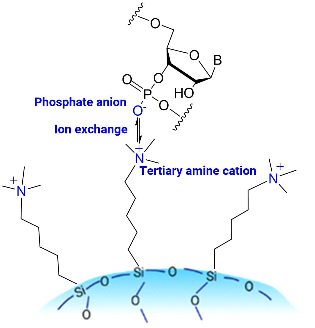
Figure 4. Schematic illustration of the anion-exchange mechanism
Technical Features and Key Influencing Factors
This method is straightforward to perform and enables automated capture, washing, and elution. It provides clean extracts and effectively addresses matrix effects. It is broadly applicable, providing satisfactory extraction from matrices such as plasma, tissue homogenates, bile, and urine.
Method development should focus on:
The choice and amount of denaturing agent, to prevent it from occupying capture sites on the beads.
Careful optimization of the capture pH: the optimal window is narrow and varies among nucleic acid therapeutics.
Optimization of the eluent: suitable alkaline conditions are required to bring the cationic groups to a neutral state and achieve effective elution.
Applicable Settings
This approach is suitable for extracting oligonucleotide therapeutics when sample numbers are large or high throughput is required, and from matrices with pronounced matrix effects, including bile, urine, and tissue homogenates.
Hydrophobic Interactions
Principle
In hydrophobic-interaction extraction, an oligonucleotide therapeutic first forms an ion pair with a basic ion-pairing reagent through electrostatic interactions. The hydrophobic moiety of the ion-pairing reagent then binds to hydrophobic groups attached to the magnetic beads through hydrophobic interactions, enabling extraction of the target oligonucleotide. The interaction mechanism is shown in Figure 5.
The extraction workflow is as follows:
Add an RNA extraction reagent (lipid-modified oligonucleotides samples require protease digestion) to the sample to dissociate siRNA from proteins.
Add a basic ion-pairing reagent at a specified concentration to form ion pairs with the oligonucleotide molecules.
Add magnetic beads bearing surface-bound hydrophobic groups to form magnetic bead–nucleic acid complexes through hydrophobic interactions.
Retain the beads by magnetic separation and wash repeatedly with wash buffer to remove unbound non-target nucleic acids and other impurities.
Elute with an organic solvent under strongly alkaline conditions to disrupt electrostatic and hydrophobic interactions and release the target nucleic acids.
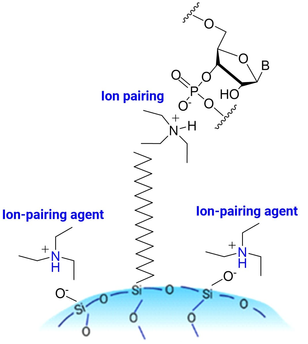
Figure 5. Schematic illustration of the hydrophobic-interaction mechanism
Technical Features
Typically, after the sample is treated with an RNA extraction reagent, the aqueous phase is collected for capture by hydrophobic magnetic beads. The principal advantage of this method is its ability to effectively eliminate matrix effects in complex matrices such as bile, urine, and tissue homogenates. In ion-exchange extraction, the addition of excess denaturing agent can reduce recovery. Hydrophobic-interaction extraction avoids this issue by directly using the aqueous phase obtained after extraction for capture. For lipid-modified oligonucleotides, the additional contribution of the hydrophobic lipid groups can improve extraction performance.
Key Influencing Factors
Method development should focus on:
Avoiding the introduction of large amounts of organic solvents or surfactants that disrupt hydrophobic interactions.
The concentration and type of basic ion-pairing reagent.
The effects of temperature on sample loading and elution.
Applicable Settings
This method is also suitable for samples with pronounced matrix effects, such as bile, urine, and tissue homogenates. It is particularly useful for tissue homogenates in which ion-exchange extraction does not provide satisfactory recovery. For lipid-modified oligonucleotides, its extraction performance is superior to that of ion-exchange extraction after protease digestion.
Application Performance and Method Selection Strategies
To directly compare the practical performance of different sample-preparation methods, the GalNAc-modified siRNA Inclisiran was used as the model drug. LLE, SPE, and three mSPE approaches— complementary base pairing, ion exchange, and hydrophobic interactions—were used to extract the target analyte from rat plasma and urine samples. The resulting extracts were analyzed under identical liquid chromatography and mass spectrometry conditions, using a Waters ACQUITY UPLC I-Class PLUS system and a SCIEX Triple QuadTM 6500+ mass spectrometer. The antisense (AS) strand ion transition 591.13/79.0 was monitored. Table 1 summarizes the comparison in terms of the lower limit of quantification (LLOQ), extraction recovery, baseline background, and the number of interfering peaks obtained with 40 μL samples.
Table 1. Comparison of five sample-preparation methods for Inclisiran in rat plasma and urine samples
Evaluation metrics | Sample type | LLE | SPE | mSPE- complementary base pairing | mSPE (Ion Exchange) | mSPE (Hydrophobic) |
Sensitivity (ng/mL) | Plasma | 10.0 | 5.00 | 2.0 | 5.00 | 5.00 |
Urine | 100.0 | 10.0 | 2.0 | 5.00 | 5.00 | |
Extraction recovery | Plasma | 71.2% | 88.6% | 71.1% | 68.7% | 68.6% |
Urine | 34.6% | 82.7% | 73.5% | 77.1% | 80.1% | |
Baseline background | Plasma | High | Medium | Low | Low | Low |
Urine | ||||||
Interfering peaks | Plasma | Many | Fewer | Fewest | Few | Few |
Urine | ||||||
Pretreatment time | Plasma | 3 h | 4 h | 5 h | 2 h | 2 h |
Evaluation metrics | Sample type | LLE | SPE | mSPE- complementary base pairing | mSPE (Ion Exchange) | mSPE (Hydrophobic) |
Plasma Sample Analysis
For plasma samples containing 10 ng/mL Inclisiran (2 ng/mL for hybridization capture), no significant differences in recovery were observed among the five methods. However, the baseline background and the number of interfering peaks differed markedly (Figures 6–10). Overall, extract cleanliness, ranked from highest to lowest (from least to most interference) was as follows:
mSPE (complementary base pairing) > mSPE (ion exchange) ≈ mSPE (hydrophobic interactions) ≈ SPE > LLE
Magnetic extraction substantially reduced baseline interference, enabling higher analytical throughput by shortening the chromatographic separation time.
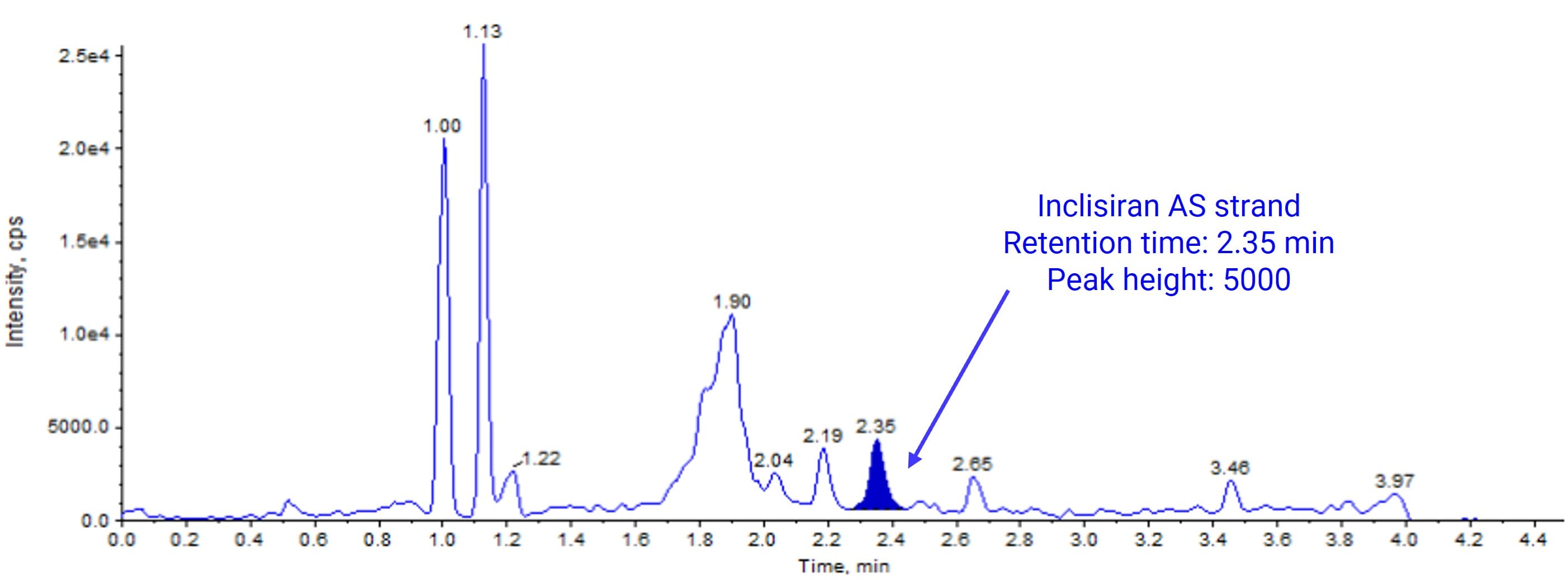
Figure 6. Chromatogram of a plasma sample spiked with 10 ng/mL Inclisiran after LLE
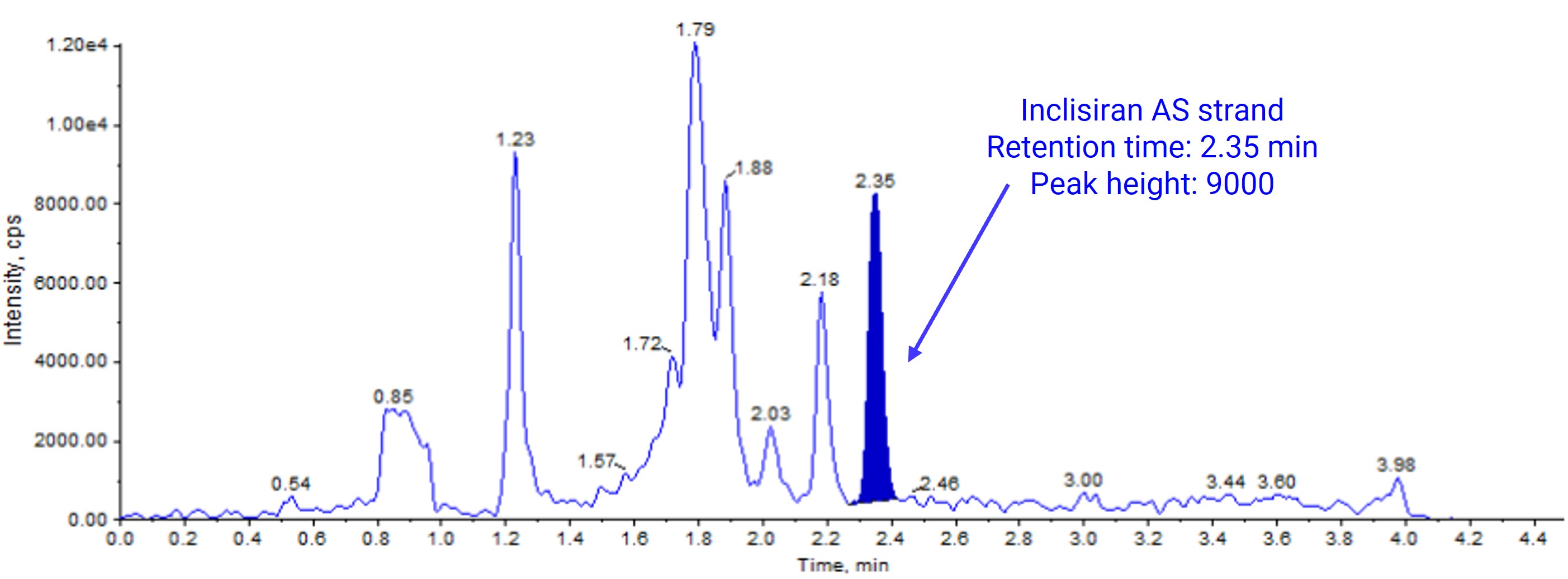
Figure 7. Chromatogram of a plasma sample spiked with 10 ng/mL Inclisiran after SPE
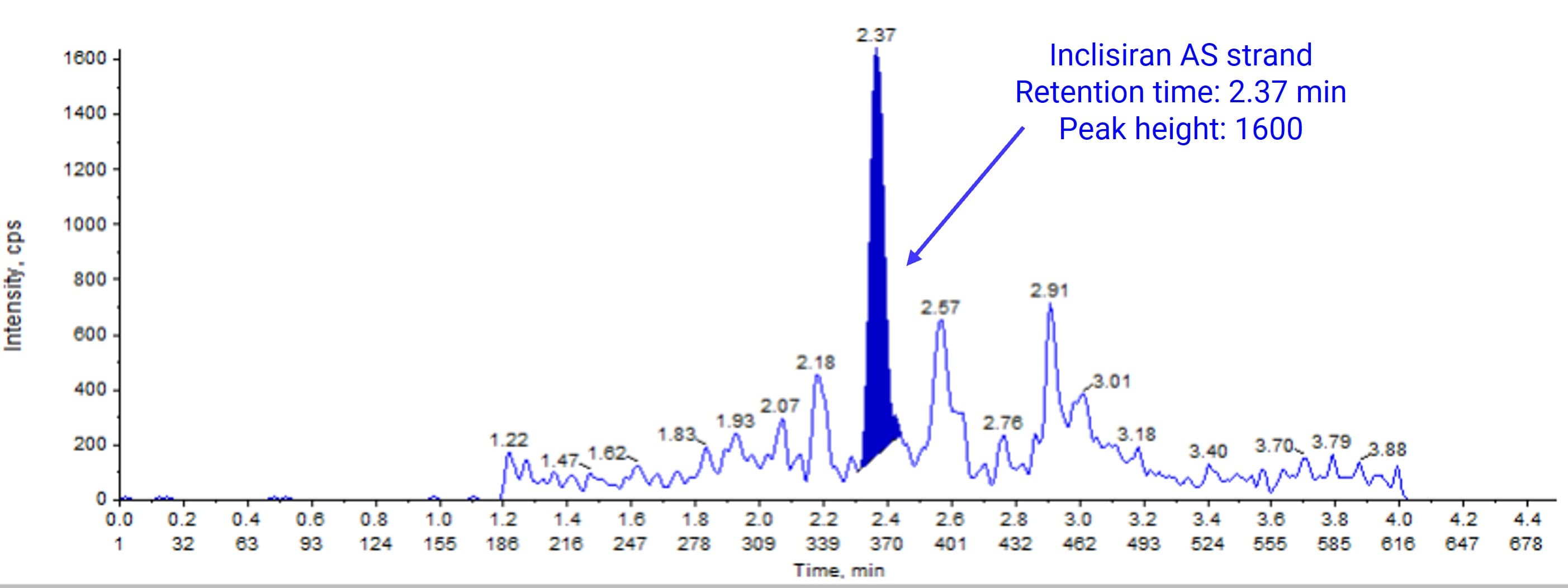
Figure 8. Chromatogram of a plasma sample spiked with 2 ng/mL Inclisiran after mSPE-complementary base pairing
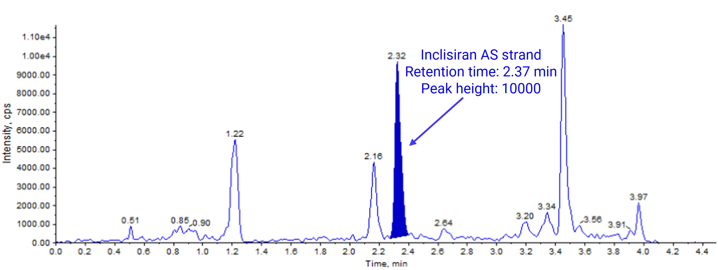
Figure 9. Chromatogram of a plasma sample spiked with 10 ng/mL Inclisiran after mSPE-ion exchange interaction
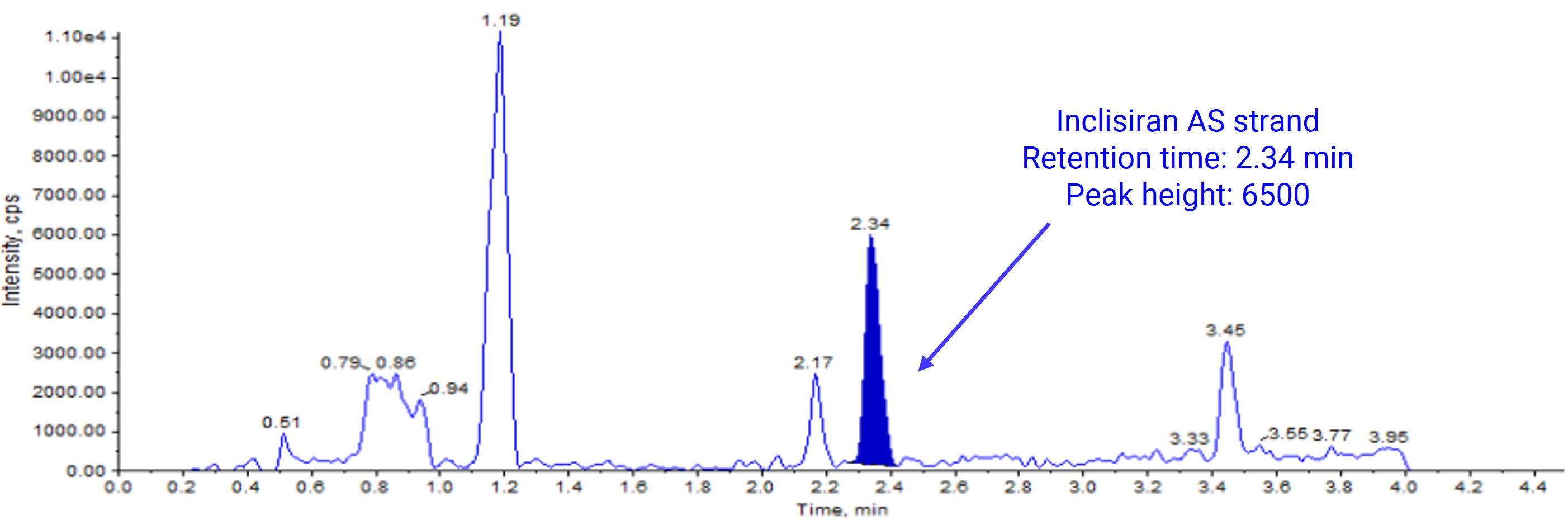
Figure 10. Chromatogram of a plasma sample spiked with 10 ng/mL Inclisiran after mSPE-hydrophobic interaction
Urine Sample Analysis
The differences among the five methods were more pronounced in urine samples spiked with Inclisiran (Figures 11–15). Recovery with LLE was only 33.6%, and the high baseline noise made it difficult to meet the analytical requirements at 10 ng/mL. SPE reduced the baseline background and achieved an LLOQ of 10 ng/mL. mSPE further reduced the background, lowering the LLOQ to 5 ng/mL. Hybridization capture provided the highest sensitivity, achieving an LLOQ of 2 ng/mL.
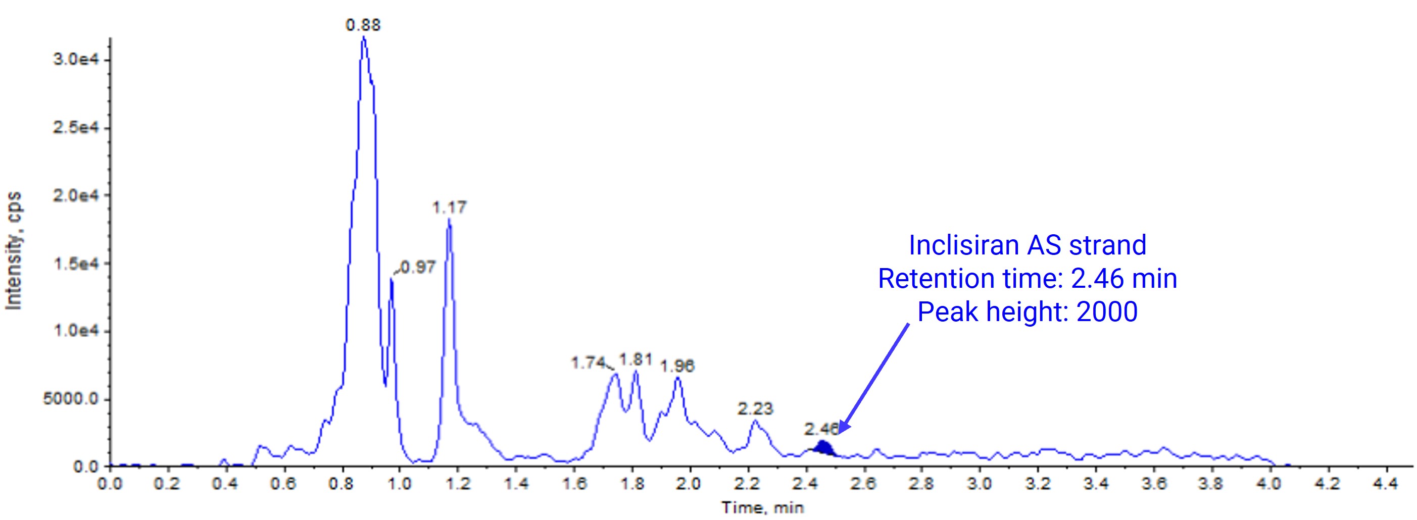
Figure 11. Chromatogram of a urine sample spiked with 10 ng/mL Inclisiran after LLE
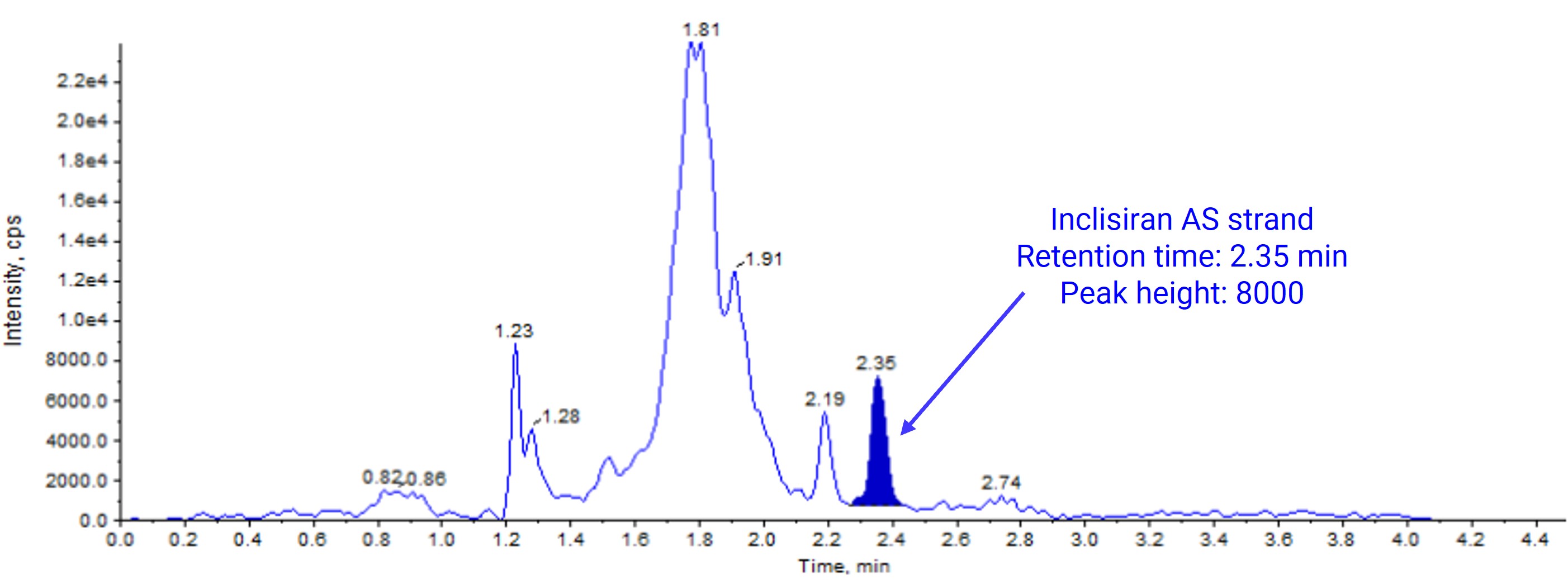
Figure 12. Chromatogram of a urine sample spiked with 10 ng/mL Inclisiran after SPE
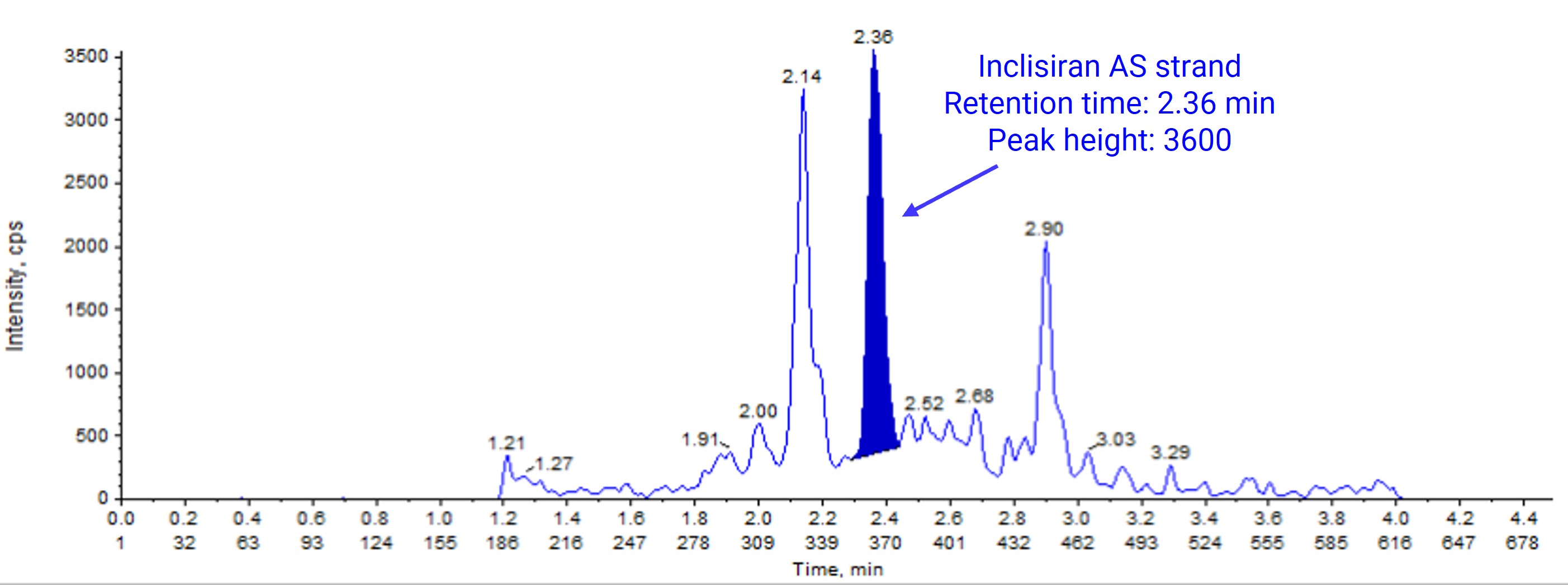
Figure 13. Chromatogram of a urine sample spiked with 2 ng/mL Inclisiran after mSPE-complementary base pairing
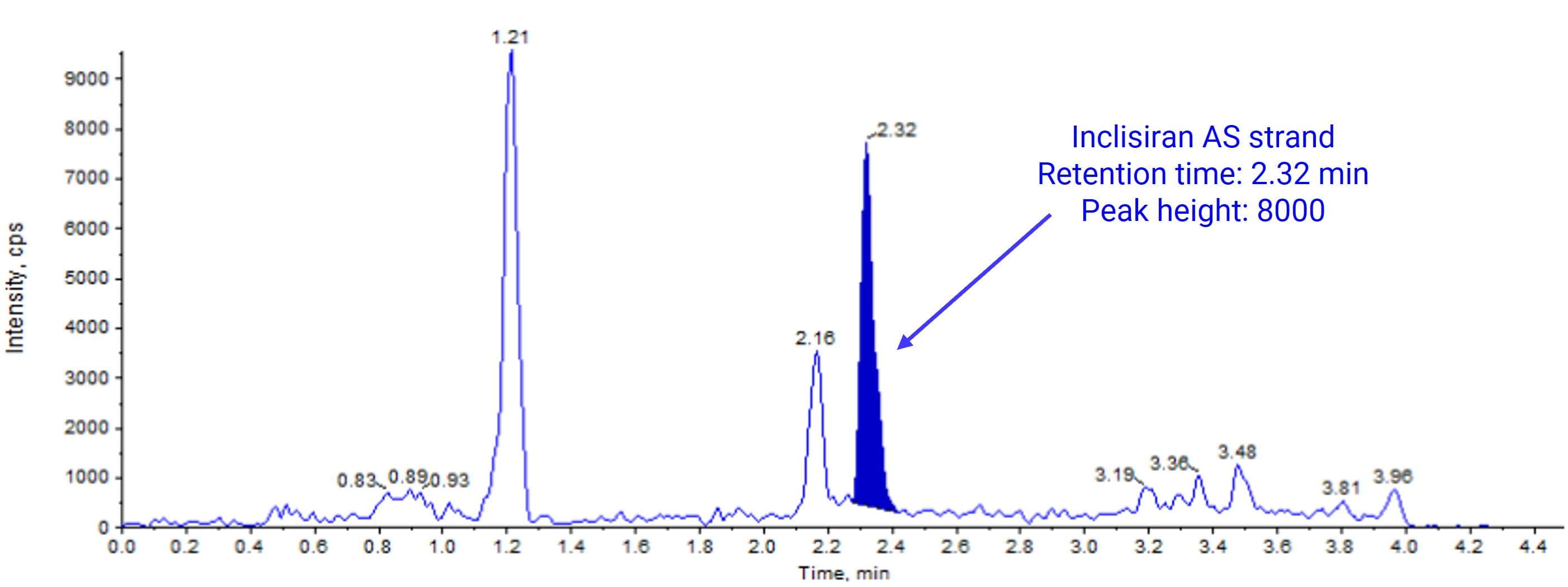
Figure 14. Chromatogram of a urine sample spiked with 10 ng/mL Inclisiran after mSPE-ion exchange interaction
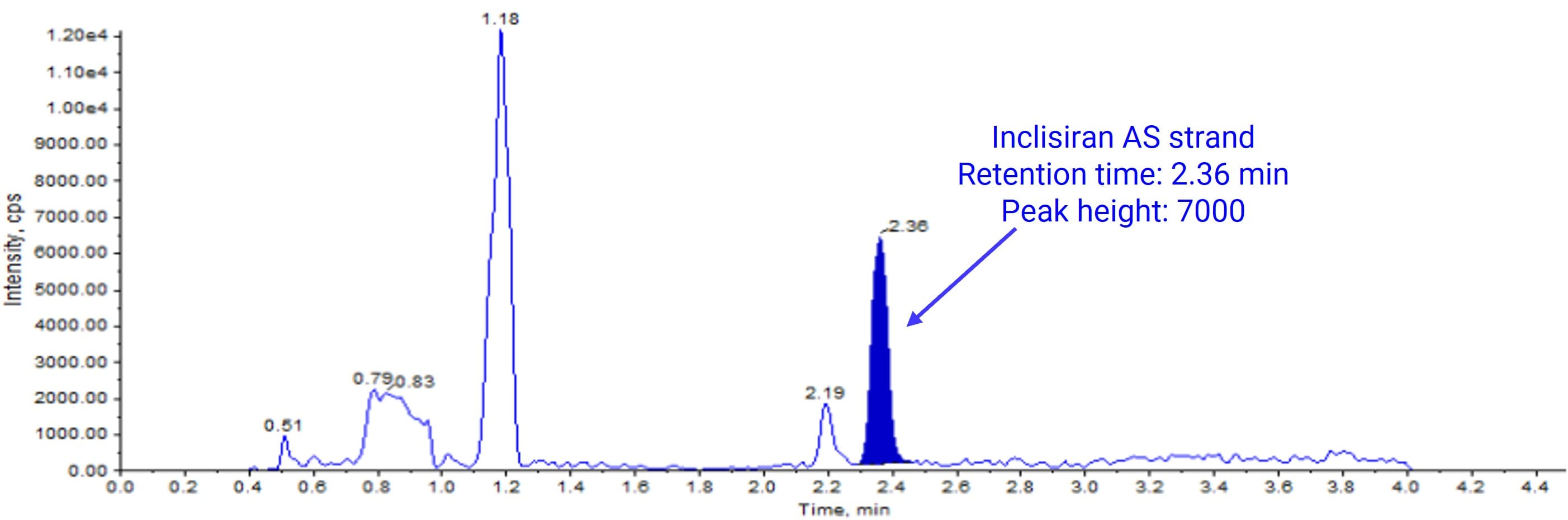
Figure 15. Chromatogram of a urine sample spiked with 10 ng/mL Inclisiran after mSPE-hydrophobic interaction
Recommendations for Method Selection
Based on these results, the applicable settings for each method can be summarized as follows:
LLE: Simple to perform and suitable for initial extraction of routine plasma samples. However, it has clear disadvantages in matrix effects, interference, and recovery when applied to matrices from tissue-distribution and excretion studies.
SPE: An established method with reliable cleanup performance that meets most bioanalytical requirements and provides a dependable alternative.
mSPE (ion exchange interaction/hydrophobic interaction): Superior to LLE and SPE in sensitivity, extract cleanliness, and operational efficiency. Particularly suitable for complex matrices, such as tissues, bile, and urine, or when higher sensitivity is required.
mSPE (complementary base pairing): The preferred option when even higher sensitivity is needed, for example for low-abundance targets or very low-dose drugs, or when the above sample-preparation methods do not meet extraction requirements. Its longer sample-preparation time must, however, be considered during method design.
Magnetic extraction not only provides excellent sample cleanup but also offers strong potential for automation. Fully automated processing from adsorption to elution can reduce human error and further improve turnaround time.
Conclusion
The bioanalysis of oligonucleotide therapeutics presents distinctive challenges for sample preparation. This article systematically compared the practical performance of conventional methods, LLE and SPE, with three modes of the emerging mSPE technology. Using Inclisiran as a case study, it demonstrated notable advantages of mSPE in sensitivity, extract cleanliness, operational efficiency, and compatibility with automation. In practice, researchers can flexibly select an appropriate sample-preparation strategy according to their analytical objectives and matrix complexity.
Drawing on extensive technical expertise and established platform capabilities in oligonucleotide bioanalysis, WuXi AppTec DMPK provides end-to-end, customized technical services, from sample-preparation method development and validation to sample analysis and data reporting, to address the specific bioanalytical needs of different stages of drug development.
Authors: Yan Song, Xiaopeng Wu, Jin Xie, Xiaoxia Yang, Peiyun An, Jinlian Lu, Lili Xing
Talk to a WuXi AppTec expert today to get the support you need to achieve your drug development goals.
Committed to accelerating drug discovery and development, we offer a full range of discovery screening, preclinical development, clinical drug metabolism, and pharmacokinetic (DMPK) platforms and services. With research facilities in the United States (New Jersey) and China (Shanghai, Suzhou, Nanjing, and Nantong), 1,300+ scientists, and over fifteen years of experience in Investigational New Drug (IND) application, our DMPK team at WuXi AppTec are serving 1,800+ global clients, and have successfully supported 2,100+ IND applications.
Reference
[1] Cheng Zhongzhe, Jiang Hongliang. Advances in bioanalytical methods for nucleic acid therapeutics. Acta Pharmaceutica Sinica. 2021;56(9):2335–2345. [In Chinese; article title translated from the supplied reference.]
[2] Sun Shumeng, Lin Lin, Zhang Daizhou, et al. Advances in bioanalytical methods for nucleic acid therapeutics based on liquid chromatographic separation. China Pharmacy. 2024;35(23):2959–2964. [In Chinese; article title translated from the supplied reference.]
[3] LIU A W, CHENG M, ZHOU Y X, et al. Bioanalysis of oligonucleotide by LC-MS: effects of ion pairing reagents and recent advances in ion-pairing-free analytical strategies[J]. Int J Mol Sci,2022,23(24):15474.
[4] Capriotti A L, Cavaliere C, La Barbera G, et al. Recent applications of magnetic solid-phase extraction for sample preparation[J]. Chromatographia, 2019, 82(8): 1251-1274.
[5] Zifeng Song, Angela Lu, Long Yuan. Analyte and probe melting temperature guided method development strategy for hybridization LC-MS/MS quantification of siRNAs[J]. J. Pharm. Biomed. Anal, 2025, 253:116556.


FAQ
-

Why is mSPE preferred over traditional solid-phase extraction (SPE) or LLE for oligonucleotide therapeutics?


-
When should hybridization capture be selected for the bioanalysis of oligonucleotides?
-
Can mSPE techniques be automated for high-throughput oligonucleotide bioanalysis?
Related Services and Platforms


-
 DMPK BioanalysisLearn More

-
Novel Drug Modalities DMPK Enabling PlatformsLearn More
-
Novel Drug Modalities BioanalysisLearn More
-
Small Molecules BioanalysisLearn More
-
Bioanalytical Instrument PlatformLearn More
-
PROTAC DMPK ServicesLearn More
-
ADC DMPK ServicesLearn More
-
Antibody-Oligonucleotide Conjugate (AOC) DMPK ServicesLearn More
-
Oligo DMPK ServicesLearn More
-
PDC DMPK ServicesLearn More
-
Peptide DMPK ServicesLearn More
-
mRNA DMPK ServicesLearn More
-
Covalent Drugs DMPK ServicesLearn More

Stay Connected
Keep up with the latest news and insights.

