



A Comprehensive Guide to Tissue Homogenization Quality Control in DMPK Studies: From Method Selection to Data Reliability
-

Articles
-

Jul 17, 2026
In drug discovery and development, tissue distribution research in preclinical pharmacokinetics (PK) characterizes the distribution dynamics changes of a drug in target and non-target organs. These studies are a critical component in evaluating drug targeting, predicting efficacy, and identifying potential tissue accumulation [1]. To quantitatively determine the concentrations of drugs and their metabolites in tissues, technologies such as liquid chromatography–tandem mass spectrometry (LC-MS/MS) have become the gold standard analytical method.
However, a crucial preprocessing step, tissue homogenization, is often overlooked. Tissue homogenization is the process of converting collected organs or tissue samples into a homogeneous liquid matrix suitable for analytical testing. The selection of homogenization methods and the control of operational details directly affect analyte stability, extraction efficiency, and the precision and accuracy of analytical results.
This article compares mainstream homogenization technologies, analyzes the differences influenced by varying sample requirements and tissue characteristics, and describes the key quality control points throughout the homogenization workflow.
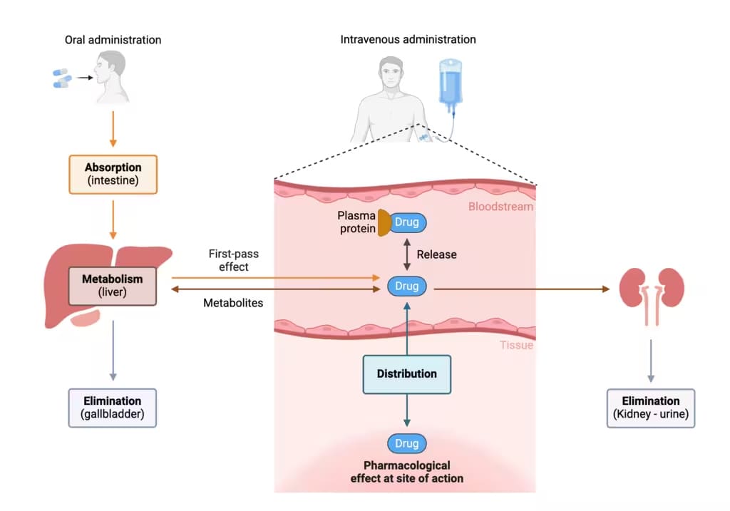
Figure 1. ADME process of drugs in vivo [1]
In general, from an analytical perspective, methods based on tissue homogenate analysis (a destructive approach for quantitative or qualitative) predominantly rely on liquid chromatography-tandem mass spectrometry (LC-MS/MS). In contrast, methods utilizing radioactivity for quantitative or qualitative analysis with spatial distribution commonly employ quantitative whole-body autoradiography (QWBA). Whereas fluorescence or chemical-based imaging methods typically employ qualitative in vivo imaging approaches.
Comparison of Homogenization Methods: Principles, Advantages, Limitations, and Application
Disruption of tissue samples is an early step in the isolation and/or quantification of RNA, DNA, proteins, and analytes. Both chemical and mechanical/physical methods can be used for tissue disruption. For biological tissue samples, mechanical disruption is generally adopted. The major considerations during sample processing include selecting an appropriate homogenization buffer, controlling temperature, using suitable homogenization equipment, avoiding contamination, and performing appropriate centrifugation. The principles and applicable scenarios of different homogenization methods are detailed in Table 1.
Table 1. Advantages, limitations, and applications of different homogenization methods
Homogenization Method | Advantages | Disadvantages | Suitable Tissue Types | Temperature Control | Potential Risks | Recommendation Index |
Manual homogenization | Simple | Low efficiency, poor reproducibility, prone to heating, difficult to process tough tissues, high contamination risk | Soft tissues (brain, liver, etc.) | Poor | High | Not recommended |
High-speed mechanical homogenization | High efficiency, rapid processing, wide applicability | Severe heat generation (requires strict temperature control), noisy, potential splashing and cross-contamination | Soft tissues (brain, liver, etc.) and hard tissues (muscle, heart, lung, tumor, etc.) | Ice bath required | Low | *** |
Bead-beating homogenization | Extremely high efficiency, low-temperature operation, high throughput, closed system | Beads may adsorb analytes; intense process may release interferents | Soft tissues (brain, liver, etc.) and hard tissues (muscle, heart, lung, tumor, etc.) | Optional (instrument-dependent) | Low | ***** |
Ultrasonic disruption | Suitable for cells/very soft tissues | Heat generation, poor effect on large tissue blocks, possible compound degradation, highly dependent on reproducibility | Cell suspensions, very soft tissues | Ice bath / intermittent operation required | Medium | ** |
Note: The recommendation index is based on a comparative assessment of the advantages and limitations of different homogenization methods.
Why is Post-Homogenization Processing Crucial for Bioanalysis?
Tissue homogenization itself is not the endpoint, but rather a necessary preparatory step for subsequent analysis techniques (such as LC-MS/MS, Enzyme-linked immunosorbent assay (ELISA), etc.). Different analytical targets and tissue sample characteristics require differentiated post-homogenization processing strategies:
LC-MS/MS: This analytical technique is highly sensitive to sample matrix effects. Incomplete removal of lipids, protein fragments, and other components generated during homogenization can suppress or enhance ionization efficiency, leading to inaccurate quantification. Therefore, the centrifugation step is critical and further sample cleanup steps (such as protein precipitation or solid-phase extraction (SPE)) are often required. The choice of homogenization buffer should be as compatible as possible with subsequent extraction solvents to minimize interference.
ELISA: As a rapid, sensitive, and accurate immunological detection method, ELISA measure a wide range of analytes/indicators. However, proteins in tissues like the liver, kidney, and brain are complex and variable. Differences in tissue fragment size/ granularity and protein extraction efficiency can lead to significant variations in protein content among samples. Consequently, for tissue sample preparation aimed at collecting target proteins, special attention must be paid to the mildness of the procedure and sample storage conditions, particularly avoiding repeated freeze–thaw cycles to minimize their impact on assay accuracy.
How to Choose the Right Tissue Homogenization Buffer?
Selecting the appropriate buffer is a key determinant in tissue homogenization strategies. Different media serve different functional purposes:
Buffers (PBS, Tris-HCl, phosphate buffers, etc.): Maintain pH stability and reduce compound degradation or shifts in ionization states caused by pH fluctuations. Specific components can be added (e.g., EDTA to chelate metal ions, sodium fluoride to inhibit enzymes).
Organic solvents (Acetonitrile, Methanol): Can rapidly precipitate proteins and inactivate enzymes, effectively quenching metabolism and degradation. This is particularly suitable for analyzing metabolically active organs (like the liver) or unstable compounds. Disadvantage: May alter tissue morphology, making the pellet more compact after centrifugation or leaving more particles in the supernatant. Centrifugation conditions need optimization, and solvent evaporation must be monitored.
Acidified solutions (e.g., water or buffers containing formic acid or trifluoroacetic acid): Help stabilize basic drugs (reducing non-specific binding to labware) and inhibit certain enzyme activities.
Buffers containing protease/phosphatase inhibitors: When the target analytes include proteins, peptides, or phosphorylated proteins, inhibitors are necessary to prevent degradation by endogenous enzymes during tissue sample preparation.
Dilution factor: The volume of medium added during homogenization determines the dilution factor of the resulting homogenate. Excessive dilution may cause drug concentrations to drop below the limit of detection (especially for tissues with low drug exposure, such as muscle). Insufficient dilution may result in a highly viscous tissue homogenate or a cloudy supernatant after centrifugation. Optimization is required based on the expected concentration range, tissue volume, and method sensitivity.
Tissue-specific Homogenization Strategies for Different Tissue Types
Biological tissues are not homogeneous materials. Different organs vary significantly in structure, composition, and biochemical properties, posing distinct challenges for homogenization, as shown in Table 2. It is therefore clear that a “one-size-fits-all” homogenization strategy is inappropriate. Homogenization must be tailored to the characteristics of each tissue type to ensure effective analyte release and stability.
Table 2. Challenges and strategies for tissue homogenization in different tissues.
Tissue Classification | Challenges | Strategies |
Highly lipophilic tissues (adipose, brain) | Adipose tissue is difficult to homogenize; drugs (especially lipophilic ones) may strongly bind to fat or the lipid bilayers of brain tissue. | Low-temperature operation is crucial (to prevent lipid melting/smearing). Add an appropriate amount of organic solvent to the homogenization medium to facilitate the release of bound drugs. |
Highly fibrotic/dense tissues (muscle, heart, skin) | Muscle fiber bundles and connective tissues make them highly resistant to mechanical disruption. | Pre-mince the tissue into smaller pieces; select higher-intensity tissue homogenization equipment. |
Enzyme-rich tissues (liver, kidney) | These organs have extremely high metabolic activity; drugs may be rapidly degraded or transformed during tissue homogenization. | Operate strictly under low temperatures (ice bath); the homogenization medium should be a buffer capable of rapidly quenching enzyme activity. |
Blood-rich tissues (liver, spleen) | Contain a large number of blood cells; hemoglobin may interfere with the analysis. | Perfusion or rinsing of the tissue with saline prior to tissue homogenization is recommended; thorough centrifugation after homogenization is required to remove cell debris. |
Hard tissues (bone) | High tissue hardness makes standard tissue homogenization methods ineffective. | Special equipment, such as a cryogenic grinder, can be used to pulverize the bone in liquid nitrogen. |
Showcase: How Does Tissue Homogenization Impact Data Reliability?
The following case demonstrates the impact of different homogenization methods on analytical results, further highlighting the importance of standardized procedures within a quality management system.
Researchers compared two automated tissue homogenization technologies with the commonly used handheld rotor homogenization method. By analyzing the degree of tissue homogenization, including particle fineness and uniformity, as well as the extraction abundance of key proteins, they comprehensively evaluated the tissue homogenization quality achieved by the three methods. The tissue types tested were liver and brain, and the homogenization buffer was 100 mM Tris-HCl at pH 7.6 [2, 3].
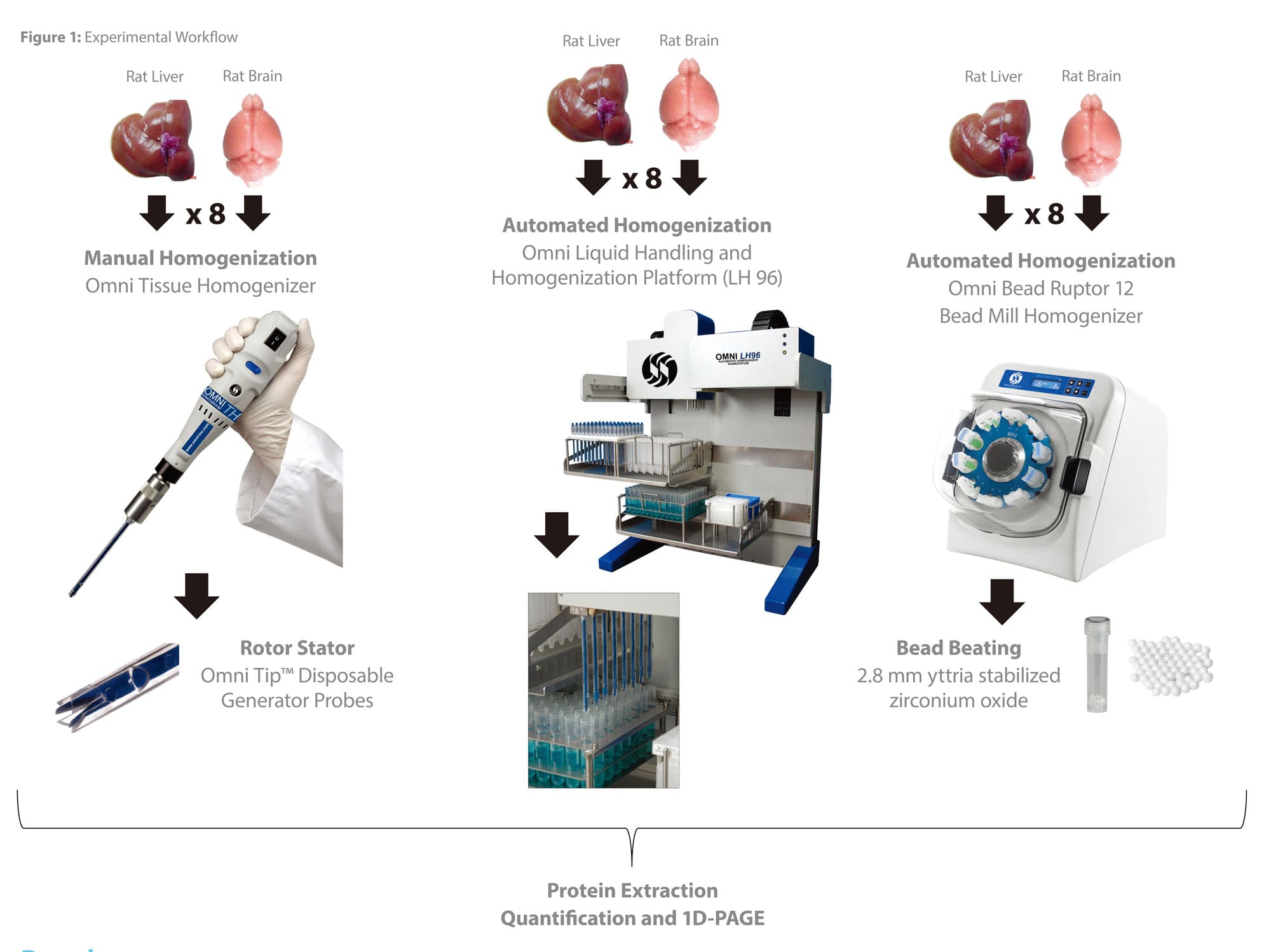
Figure 2. Experimental workflow for tissue homogenization operations[3]
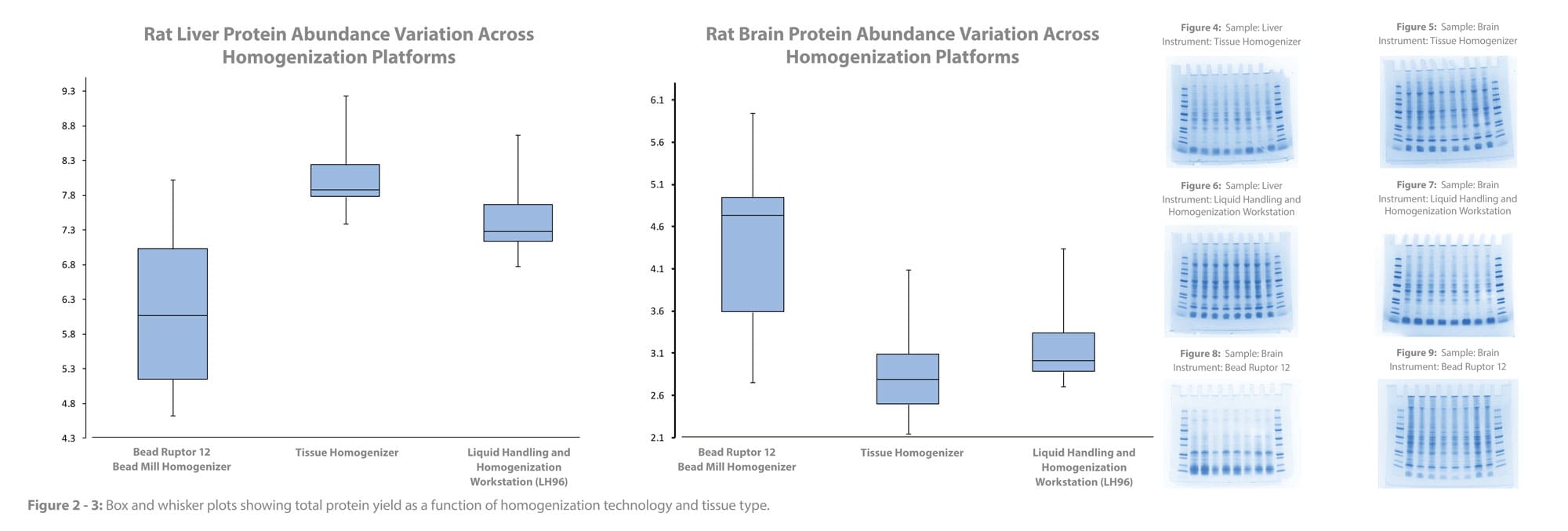
Figure 3. Total protein content and protein abundance analysis after homogenization using different instruments [3]
The experimental results showed that all three methods produced acceptable total protein extraction yields. However, comparison of protein abundance measurements revealed significant differences in the extraction yield of the same protein across different methods. Studies involving protein, nucleic acid, and small-molecule analysis typically begin with homogenization to release target analytes. An increasing body of data indicates that experimental variability and analytical sensitivity depend to a large extent on differences in analyte extraction efficiency during the homogenization process.
Conclusion
Tissue homogenization serves as the critical bridge between in vivo drug distribution phenomena and in vitro quantitative measurement. The quality of this process directly determines whether the data observed by researchers truly reflect the actual distribution of a drug during DMPK studies.
This article systematically reviewed the advantages, limitations and application of mainstream homogenization technologies. It emphasized the necessity of sample-specific approaches, tailored sample processing based on analytical goals (drug/metabolite stability, analytical technology requirements) and tissue characteristics (lipophilicity, fibrosis, enzyme content). Furthermore, it detailed key quality control points such as temperature control, buffer selection, equipment selection, contamination prevention, and centrifugation conditions during the preparation of a tissue homogenate.
At WuXi AppTec DMPK, service quality and execution excellence form the cornerstones of our operations. Through extensive practical experience in DMPK studies, we have established standardized homogenization SOPs (Standard Operating Procedures) through a rigorous quality control system. Our process prioritizes optimal tissue homogenization methods tailored to different tissue types and analytical goals, with continuous monitoring of key parameters (temperature, buffer, and centrifugation). This ensures the generation of robust and reproducible data from every tissue sample, thereby delivering a trustworthy foundation for our clients' drug R&D decision-making.
Authors: Furong Jiao, Cheng Tang
Talk to a WuXi AppTec expert today to get the support you need to achieve your drug development goals.
Committed to accelerating drug discovery and development, we offer a full range of discovery screening, preclinical development, clinical drug metabolism, and pharmacokinetic (DMPK) platforms and services. With research facilities in the United States (New Jersey) and China (Shanghai, Suzhou, Nanjing, and Nantong), 1,000+ scientists, and over fifteen years of experience in Investigational New Drug (IND) application, our DMPK team at WuXi AppTec are serving 1,600+ global clients, and have successfully supported 1,800+ IND applications.
Reference
[1] Pharmacokinetics (ADME): an overview. http://pharmacologymentor.com/?s=Pharmacokinetics+%28ADME%29%3A+an+overview.
[2] Piehowski PD, Petyuk VA, Orton DJ, Xie F, Moore RJ, Ramirez-Restrepo M, Engel A, Lieberman AP, Albin RL, Camp DG, Smith RD, Myers AJ. Sources of technical variability in quantitative LC-MS proteomics: human brain tissue sample analysis. J Proteome Res. 2013 May 3;12(5):2128-37.
[3] Evaluating the Efficacy and Reproducibility of Automated Homogenization Technologies.


ON THIS PAGE

Related Resources


Related Services and Platforms







Stay Connected
Keep up with the latest news and insights.

