



Targeted Release Principle and Metabolite Identification (MetID) Study of Peptide-Drug Conjugates (PDCs)
-

Articles
-

Oct 23, 2026
An ideal drug delivery system enhances efficacy, broadens the therapeutic window, reduces the dosage, and minimizes the toxic and side effects through targeted delivery and controlled release. Particularly in the field of cancer therapy, the precise and efficient killing achieved by targeted delivery drugs has significant advantages over the indiscriminate damage caused by radiotherapy or chemotherapy to both tumor cells and normal cells (Figure 1).
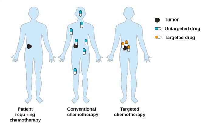
Figure 1. Tumor non-targeted and targeted therapy[1]
By the end of 2022, 15 antibody-drug conjugates (ADCs) utilizing antibodies as targeted delivery systems had been approved for marketing worldwide. Peptide-drug conjugates (PDCs), which are also emerging as a new modality, are composed of peptides as drug delivery carriers and small molecule compounds as payloads connected through linkers. Compared with ADCs, a peptide-drug conjugate has a smaller molecular mass (generally < 8 kDa), features low immunogenicity, adjustable physical and chemical properties, and low manufacturing costs.
Biologics are generally not metabolized by CYP450 enzymes but are mainly cleared through glomerular filtration (< 60 kDa), enzymatic hydrolysis, and catabolism. Compared with ADCs, PDCs with a relatively small molecular weight are cleared more rapidly with a shorter plasma exposure time. Nevertheless, targeting remains one of the most important design principles in PDC drug development from a safety and efficacy perspective. Meanwhile, conducting comprehensive metabolite identification (MetID) is essential for evaluating drug release and targeting efficiency in peptide-drug conjugates.
What Are the Metabolic Characteristics of Marketed Peptide Drug Conjugates?
The approved peptide-drug conjugate Pepaxto®(melphalan flufenamide) improves bioavailability by coupling the toxin molecule melphalan with a fluorobenzamide through peptide bonds to improve lipophilicity. Metabolic release of toxin molecules occurs through passive transport (active transport of non-protein targets) into the cell, followed by hydrolysis by aminopeptidases which is highly expressed in tumor cells (Figure 2)[2a]. The design concept here is more similar to that of a prodrug in this case.
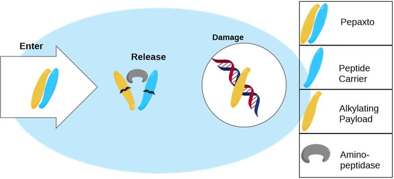
Figure 2. Mechanism of Action (MOA) of Pepaxto®[2b]
Another class of marketed PDCs is radionuclide drug conjugates (RDCs) using peptides as targeting ligands. For example, Lutathera®[3] is a drug composed of octreotide coupled with the radionuclide 177Lu. Taking advantage of the targeting of octreotide to the SSTR2 receptor, targeted radiotherapy of radionuclides can be achieved without the need to release the loaded nuclide through metabolism after being captured by tumor cells. Lutathera is not metabolized by the liver and is mainly cleared by the kidney (Figure 3).
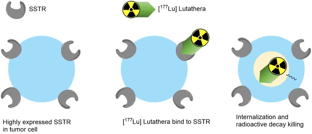
Figure 3. MOA of Lutathera®[3]
Design Principle for Targeted Release in Peptide-Drug Conjugates
At present, most of the mainstream PDC in the clinical/preclinical stage use chemotherapeutic drugs and toxin molecules (cellular microtubule inhibitors or nucleic acid inhibitors such as paclitaxel, camptothecin, maytansine, aplysia toxins) as payloads. Macromolecules in the coupled state have steric inhibition, and their payloads need to be released by metabolic degradation before they can be effective. Similar to ADCs, it is expected to maintain structural integrity and stability in the circulatory system for PDCs before entering the target cells, while they can effectively release the payloads after entering the target cells. The design idea is as follows:
The effective delivery of the payload depends on the targeted design of the peptide.
The precise release of the payload depends on the targeted release mechanism.
Targeted Delivery: Target Receptor versus Targeted Peptide
Targeted peptides targeting specific receptors on tumor cells can be obtained by receptor target screening using phage display technology or other approaches, and common receptors on tumor cells are shown in Figure 4.
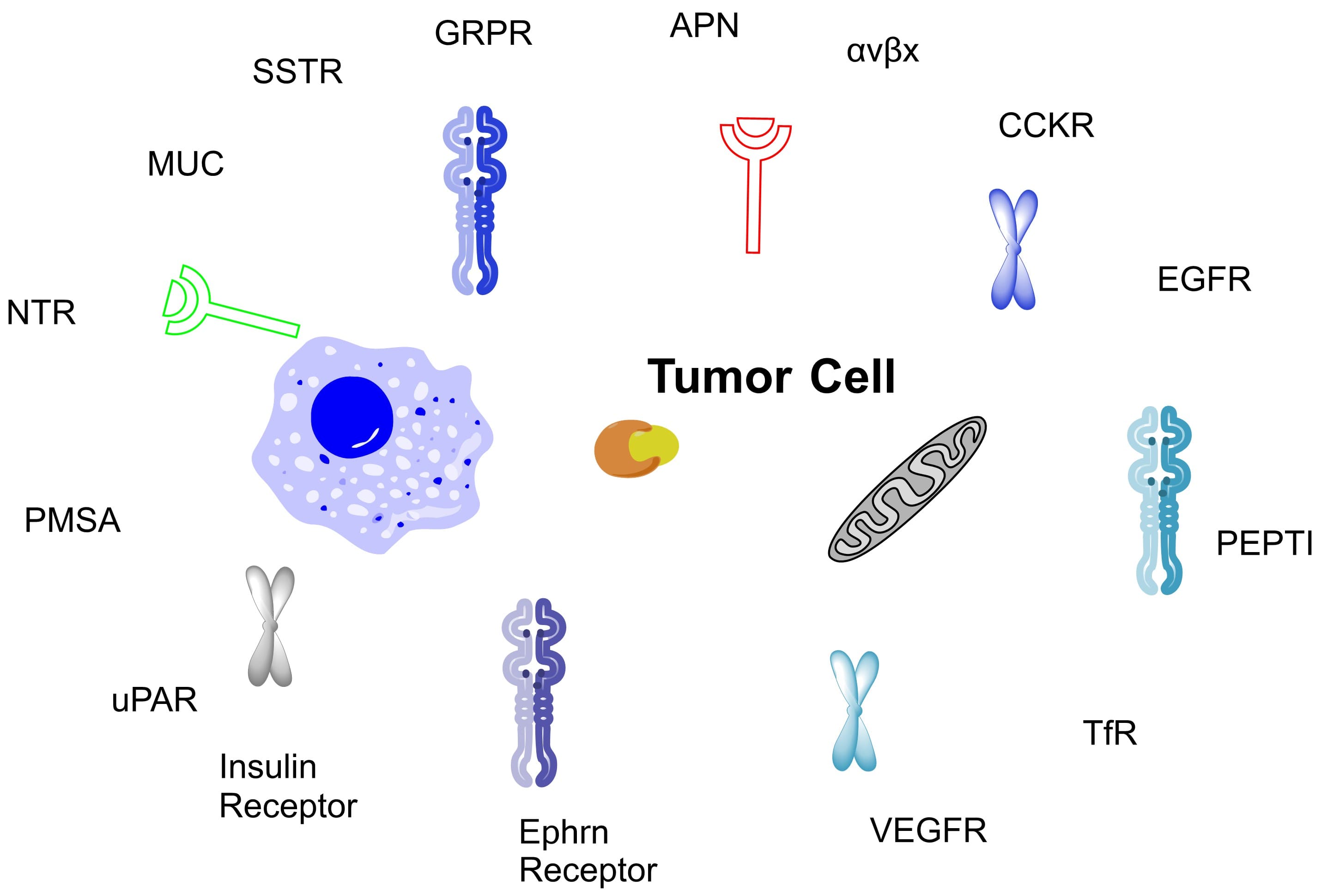
Figure 4. Common receptors on tumor cells[4]
Table 1. Partial targets and targeted peptide sequences[4]
Receptors | Targeted peptide sequence |
αvβ3 | Cilengitide [cyclo (RGDf-N(Me)V-)] |
αvβ6 | RTDLXXL |
APN/CD13 | NGR |
PEPT | Ser-Glu |
EGFR | GE11 (YHWYGYTPQNVI) |
HER2 | KCCYSL |
PSMA | KYLAYPDSVHIW |
uPAR | VSNKYFSNIHW |
GRPR | EQRLGNQWAVGHLM |
SSTRs | OCT (FCFWKTCT) |
VEGFR | Peptide SP5.2 (NGYEIEWYSWVTHGMT) |
Insulin | MCR (RRLFYKKVGLFYKKVRR) |
Ephrin receptors | EWLSPNLAPSV |
Targeted Release: Payload Release Mechanism
After PDCs are targeted into tumor cells, there can be different payload release methods according to different designs of linkers.
Non-cleavable linkers: The proteolytic enzymes of lysosomes release the payload after degradation of the peptide moiety.
Cleavable linkers: The breakage of linkers occurs in pathways such as redox, and hydrolysis (including hydrolysis in lysosomes) in the microenvironment of tumor cells, thereby releasing payloads (Figure 5).
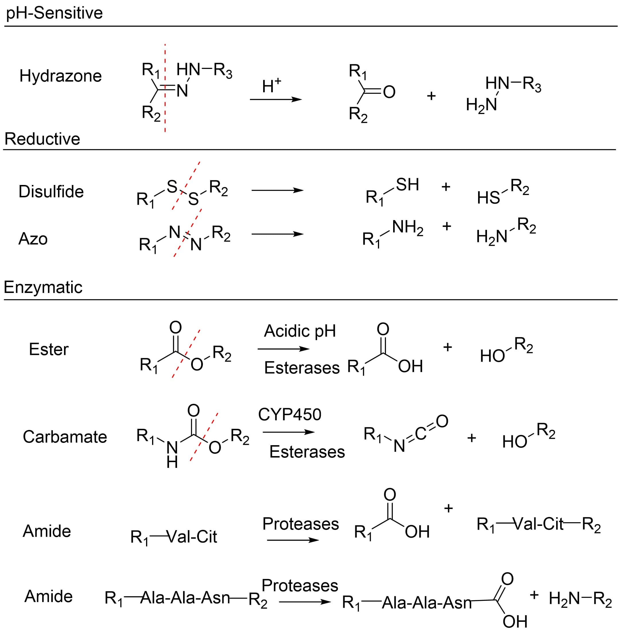
Figure 5. Common linker-cleavage mechanism[5]
Acid-cleavable linkers utilize the pH difference between tumor cell lysosomes (pH<5) and plasma (pH 7.4), selectively metabolizing within tumor cells to release the active payload, such as hydrazone that can be decomposed into aldehydes/ketones and amines under acidic conditions.
Disulfide linkers can be released by glutathione-mediated disulfide reduction/exchange to form cleavage, and the glutathione content in tumor cells exceeds that in blood by 1,000 times. Azo linkers can be cleaved under the catalysis of reduced coenzyme II (NADPH)-azoreductase, which is overexpressed in the hypoxic environment of tumor cells.
Cathepsin B is a proteolytic enzyme highly expressed in tumor cells/tissues. Cathepsin B could recognize hydrolytic specific peptide sequences, such as valine-citrulline (Val-Cit) and GFLG, and these short peptides can be used as directed enzyme cleavable linkers. In addition, when the enzyme cleavable linker is used, the self-degrading group is often used at the same time. For example, a para-aminobenzyl carbamate (PABC) linkage was used as a self-immolative spacer. When the linker is hydrolyzed, the self-immolation group undergoes spontaneous chemical elimination to release the payload (Figure 6).
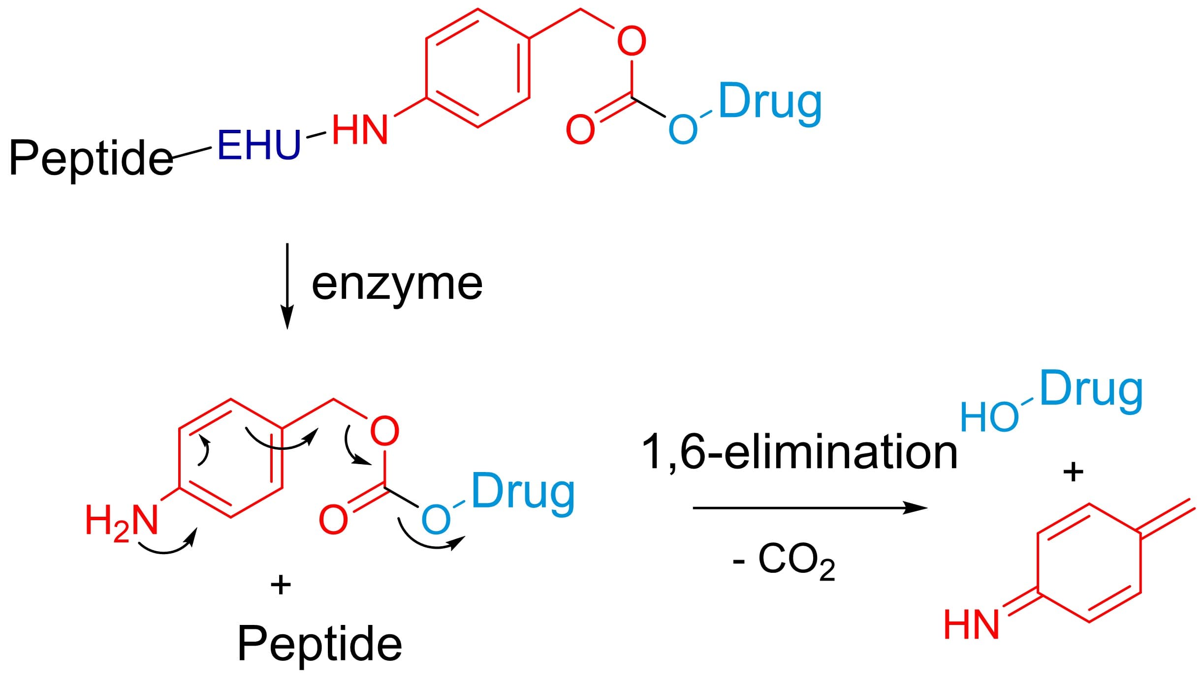
Figure 6. Mechanism of digestion - elimination of self-degradable groups[1]
Examples of PDCs in the Clinical Stage
At present, the mainstream companies that focus on PDC include Angiochem, Bicycle Therapeutics, Cybrexa Therapeutics, Oncopeptides, PeptiDream, Theratechnologies, and Borui Pharmaceutical. The following are some PDCs that have entered the clinical stage, which are composed of different peptides, linkers, and payloads, and their targeting properties and release mechanisms are typically representative.
AEZS-108[6]: Adriamycin (doxorubicin) is used as the payload and LHRH peptide is used as the targeting peptide, which is catabolized and released under the action of lysosomes after entering the cell. It has entered phase III clinical stage.
ANG1005[7]: It contains peptide-coupled multiple paclitaxel targeting the low-density lipoprotein LRP-1 receptor, which is highly expressed on tumor cells and mediates drug transmembrane entry into tumor cells, followed by release of paclitaxel through hydrolysis in lysosomes. It has entered clinical phase II/III.
CBX-12[8]: It is an antigen-independent targeted drug composed of pHLIP® peptide (in cooperation with pHLIP, Inc.), linker and small molecule anticancer drug based on the alphalexTM platform developed by Cybrexa Therapeutics, Inc. The principle is to target the specific low-pH environment of the tumor which could form alpha helix, allowing for the insertion of the peptide within the cell membrane. After delivery of the C-terminal toxin across the membrane, the payload would be released through the cleavage of disulfide bonds. CBX-12, as a candidate lead compound, has entered clinical phase I/II.
Bicyclic peptide series PDC[9]: Bicyclic peptides, developed by Bicycle Therapeutics, Inc., have high targeting, stability, and affinity. BT1718 is composed of the cleavable disulfide-linked payload DM1 and bicyclic peptides while BT8009 and BT5528 use MMAE as the payload and the cleavable dipeptide Val-Cit as the linker (Figure 7), which has entered clinical phase I/II status.
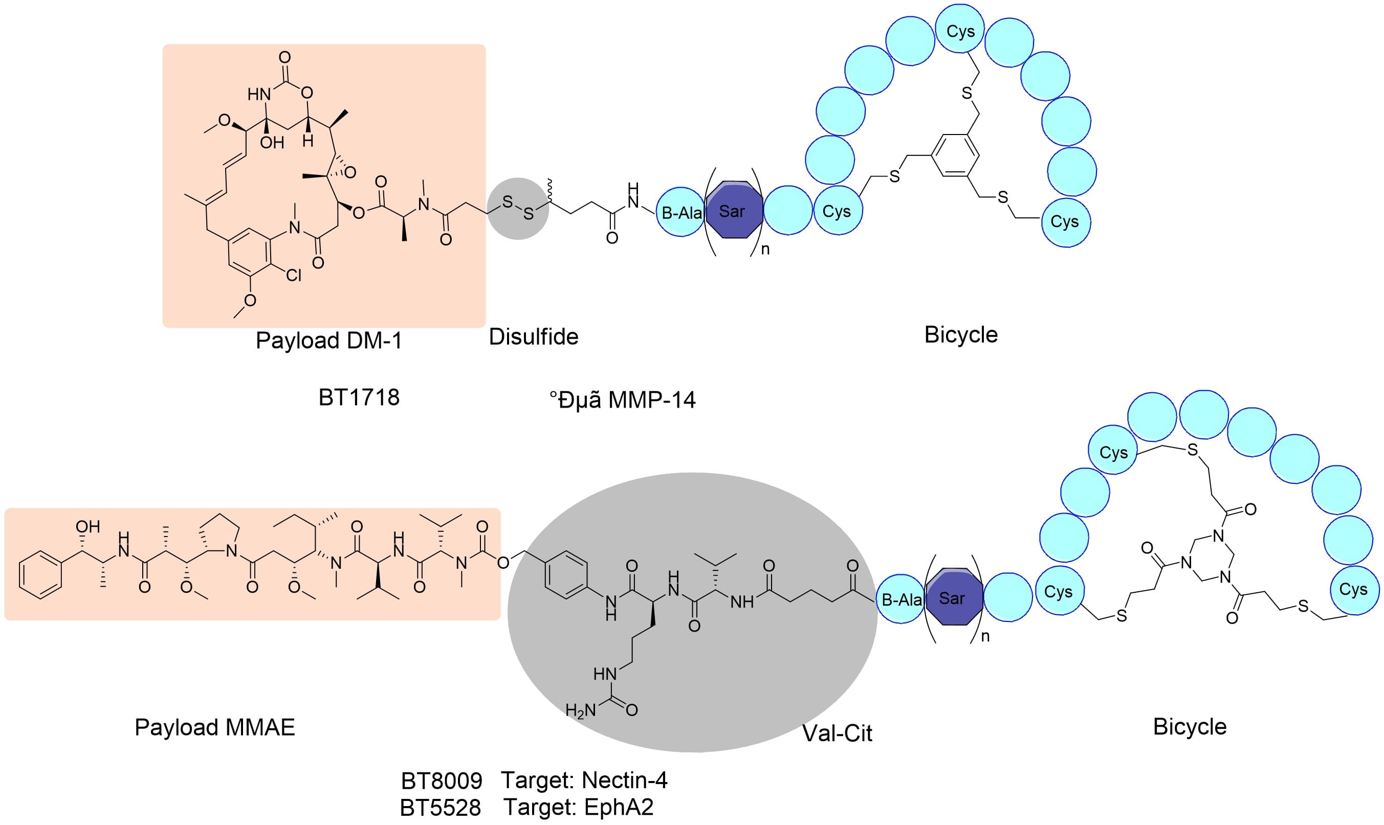
Figure 7. Structure of BT1718, BT8009, and BT5528. Adapted from reference[9]
Other Research Hotspots of PDC
Using the combination of different functional peptides and multiple site-cleavage patterns, the advantages of multiple structures can be combined from functionality and targeting. Deng[10] and others designed a multifunctional peptide combinatorial PDC consisting of targeting peptides (TTP), linked peptides, cell-penetrating peptides (CPP), and toxin molecules, targeting the LHRH receptor of tumor cells with LHRH peptides, using peptides (PLGLAG) that can be specifically degraded by MMP2 enzymes highly expressed between tumor tissues as the linker arm. After degradation of the linker peptide, toxin molecules are introduced into the cell along with the transmembrane peptide, and toxin molecules are released by rapid hydrolysis of the penetrating peptide or cleavage of the cleavable linker (Figure 8). The design simultaneously uses targeted peptides that promote the accumulation of drugs in tumor cells and transmembrane peptides that facilitate transport into cells, as well as two environmentally targeted lysis mechanisms, intracellular and extracellular, to explore new targeting and improve pharmaceutical properties.
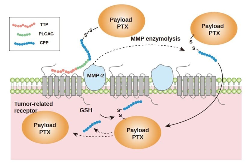
Figure 8. PDC of Targeted Peptide (TTP) - Linked Peptide - Penetrating Peptide - Toxin Molecules[10]
Metabolite Identification (MetID) Strategies and Case Studies for Peptide Drug Conjugates
In Vitro and In Vivo Metabolism Research System of PDCs
At the early stage of drug development, in vitro metabolism research systems, such as acidified liver S9 (simulating lysosomal degradation in an acidic environment of tumor cells), plasma/blood, hepatocyte, and kidney S9, can help to determine the stability and targeting of PDC, as well as the release mechanism of payload. Distinguishing the actual form of payload-related products released by PDC as early as possible can help optimize and improve the structural design and can also provide accurate information on expected metabolites for subsequent pharmacokinetics and toxicokinetics studies. Preclinical and clinical in vivo studies, on the other hand, confirmed the metabolism of PDCs and released payloads in vivo by MetID in plasma and excreta, such as urine (Table 2, Table 3).
Table 2. In vitro metabolism study system and purpose of PDCs and their payloads
In vitro test article | Incubation system | Purpose |
PDC | Plasma/blood and hepatocytes | To study the stability and off-target possibility in its circulatory system |
Acidified liver S9/tumor cells | To evaluate the payload release and analysis of its associated metabolite | |
Kidney S9 | To assess the possible metabolic pathway of payload during its renal clearance | |
Payload | Hepatocytes/microsomes/plasma | Metabolic pathways to study payload and metabolic enzymes |
Table 3. Purpose of in vivo metabolism for PDC
In vivo test article | Sampling matrix | Purpose |
PDC | Plasma/blood | To assess the safety by confirming the exposure of payloads and their associated metabolites in the circulatory system |
Urine/feces | To identify the routes of elimination for excretion and assess recovery | |
Tissue homogenate | To investigate the targeting or accumulation of drugs or metabolites in vivo |
Like conventional small molecules, the payload itself, or the identified main release products of PDC (such as payload-linkers and other main released moieties), assessment of metabolism is also required to determine its possible metabolic pathway (Figure 9, the metabolic pathways of commonly used payload MMAE), which is then helpful for the related drug interactions and toxicology studies.
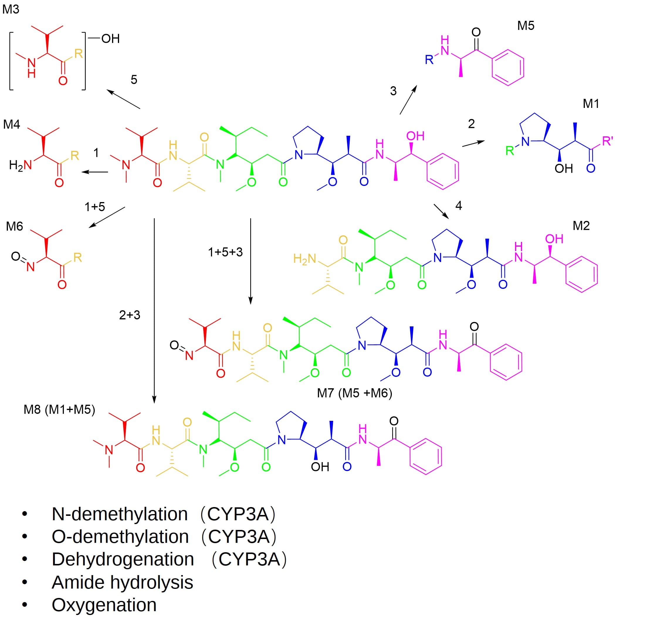
Figure 9. Metabolites and metabolic pathways of MMAE in humans[11]
Experimental Method for MetID of PDC
The MetID of PDC mainly employed a liquid chromatography-Ultraviolet-High Resolution Mass Spectrometer (LC-UV-HRMS). The analytical samples obtained by incubation with the above-mentioned in vitro study system or collected in vivo samples are treated with protein precipitation. After centrifuging, the supernatant is analyzed by LC-UV-HRMS for metabolite profiling. If the compounds are chemically stable during N2 drying, they can also be analyzed after concentration. If the peptide is neutral, the reverse liquid chromatographic system with water/acetonitrile can be used for gradient elution. If the peptide is strongly alkaline (such as transmembrane peptide TAT and other peptides containing more basic amino acid residues), an appropriate amount of trifluoroacetic acid can also be added to the mobile phase to improve the peak shape.
The components were separated by liquid chromatography and the chromatographic peaks of the metabolites were found. Then the m/z information of the mass spectrum was compared with that of the parent drug. The metabolic changes and metabolic sites were analyzed according to the primary mass spectrum (MS) and secondary mass spectrum (MS2) signals of the high-resolution mass spectrum.
Challenges and Solutions in MetID and Metabolite Profiling
The analysis of metabolites by liquid chromatography-tandem mass spectrometry (LC-MS/MS) focuses on chromatographic tracer localization and structure elucidation. Radioisotopic and non-isotopic methods are used for chromatographic tracer localization. The radioisotopic method uses radiolabeled compounds for metabolic experiments, but the synthesis of radiolabeled compounds is complicated with high costs and laboratory qualification requirements are high, which sets obstacles in the screening stage. Non-isotopic methods typically rely on additional peaks compared to the blank matrix for tracer localization, such as LC-UV or LC-MS/MS in MetID studies.
Due to their structural characteristics, PDC and its main metabolites may have weak UV absorption, so UV detection cannot effectively trace and locate them. Hence, it is necessary to obtain the secondary chromatography by data processing methods such as LC-MS/MS background subtraction to locate the possible exogenous new peaks on the chromatography (as shown in Figure 10 and Figure 11, D-E-F in the following cases).
Compound molecular size can affect the complexity of structure elucidation in MetID. Similar to ADCs, due to the steric hindrance, their payloads are usually metabolized by CYP enzymes after targeted release. However, generally, only the release mode and metabolites of payloads are considered for ADCs. The antibody part is mainly degraded through the catabolic mechanism in the body, while the peptide and linker of PDCs may be metabolized by hydrolases. Therefore, when analyzing the possible metabolites and judging the sites of metabolic changes, the payload, the peptide, and the linker need to be taken into consideration. At present, the commonly used data processing software primarily utilizes mass changes of metabolic characteristics for library matching and combined with exhaustive matching based on chemical bond breakage. Therefore, both manual analysis and software calculation are used in the data processing of MetID for PDCs. With the increase of molecular weight (number of chemical bonds), its computational complexity increases by geometric orders compared with small molecular compounds. Therefore, upgrading the software and algorithm is important to improve the efficiency of MetID for PDCs.
Case Study: In Vitro Metabolite Identification (MetID) of PDC
The following is a case study based on in vitro metabolism of a PDC with cyclic peptide and a cleavable linker. The instrument used is shown in Figure 13.
The metabolism of this PDC in acidified liver S9 (pH = 5) and hepatocyte incubation systems was compared. In the acidified S9 incubation system, only the UV peaks of the parent drug and one metabolite M1 could be found in the LC-UV spectrum, and the mass spectrum peak of another major metabolite M2 could be found by background subtraction (sample minus blank matrix) in the LC-MS/MS during MetID (Figure 11). By comparing the signals of MS and MS2, it was confirmed that the cyclic peptide M1 and the payload M2 were obtained after hydrolysis of the linker. Because the characteristic UV absorption is mainly contributed by some amino acids of cyclic peptides, the UV absorption of M2 is weak and not effectively detected on LC-UV, which can only be detected by LC-MS/MS identification during metabolite profiling.
In the hepatocyte incubation system, since the metabolite level was low, no significant metabolite peak was found in the LC-UV, and two low-abundancy metabolites M3 and M4 were found after background subtraction processing in the LC-MS/MS (Figure 12). By comparing the signals of MS and MS2, it was judged that the payload-linker part did not change, while the metabolic change was mainly the hydrolysis of the peptide part. The molecular weight and mass changes of each amino acid residue were compared, and the generated peptide after hydrolysis was concluded to finalize the metabolite identification.
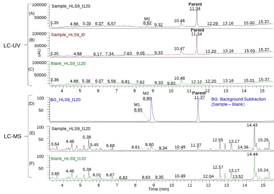
Figure 10. LC-UV (A: 120 min sample; B: 0 min sample; C: blank sample) and LC-MS/MS spectrum (D: after background subtraction; E: 120 min sample; F: blank sample) of a PDC after incubation with human acidified liver S9 system.
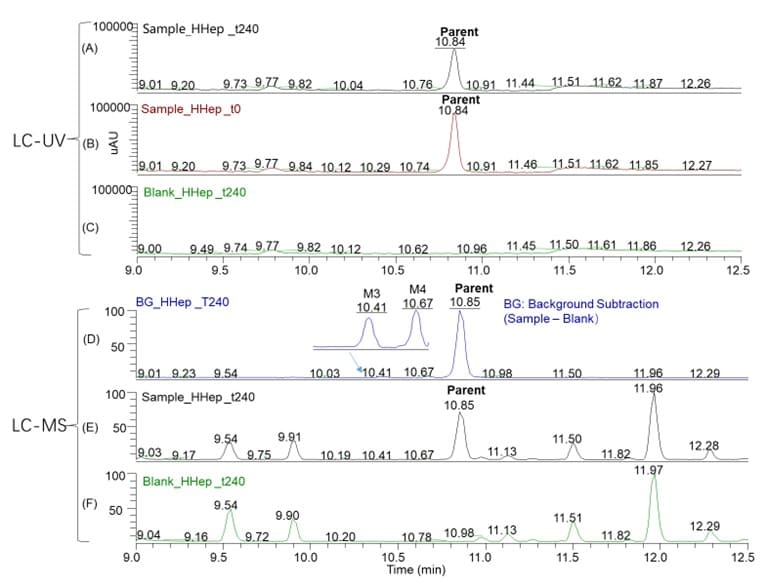
Figure 11. LC-UV (A: 240 min sample; B: 0 min sample; C: blank sample) and LC-MS/MS spectrum (D: after background subtraction; E: 240 min sample; F: blank sample) of a PDC after incubation with acidified S9 and hepatocytes.
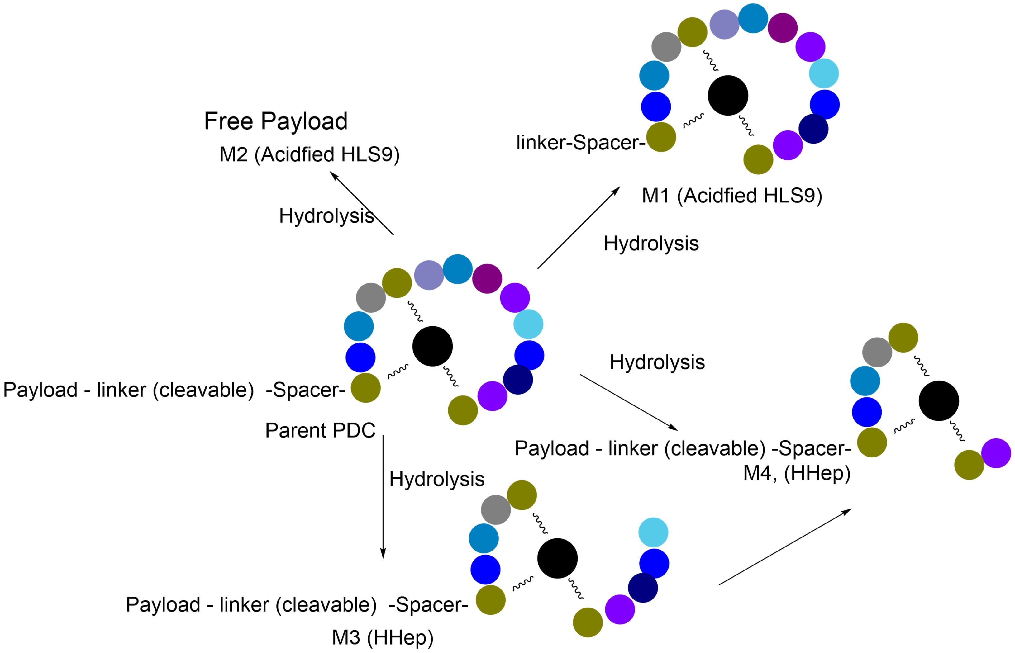
Figure 12. Difference in metabolic pathways of a PDC after incubation with acidified S9 and hepatocytes
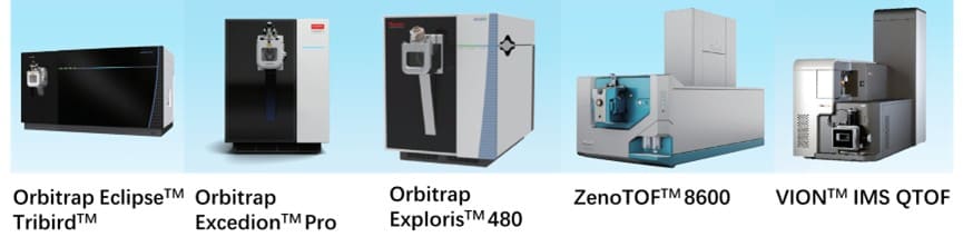
Figure 13. High-resolution LC-MS/MS analysis and product structure elucidation for identification of PDC metabolites[12]
This PDC was found to be mainly hydrolyzed through linkers to achieve targeted release of payload M2 and cyclic peptide M1 in acidified S9; partial hydrolysis on cyclic peptides occurred in the mass spectrum to produce M3 and M4, while linkers remained stable. From the difference in metabolic pathways (Figure 12), it can be seen that the metabolic release mode of payload also has tumor environmental targeting (relative to hepatic metabolism).
In addition, Val-Cit-type linkers commonly used in conjugated drugs are unstable in mouse plasma, which is due to high expression of Ces1c enzyme in mouse plasma that can hydrolyze the linkers, while it is very low in human plasma, so there will be a large variation when using mice as an animal model to study the pharmacokinetics, which needs more attention. In the study of such compounds, we also confirmed that the free payload generated in the mouse plasma is much higher than that generated in the human plasma by the in vitro plasma incubation system.
Conclusion: Advancing MetID for Peptide Drug Conjugates
With the innovative development of peptide and linker technology, PDCs have a broad development prospect. Metabolic studies of PDCs simultaneously include assessments of the metabolic stability, targeting, and payload release. This section summarizes the metabolic characteristics and research strategies of PDCs, and a MetID case is shared. More PDCs are expected to be designed and developed to bring good news to patients, supported by robust MetID and metabolite profiling platforms.
Authors: Liqi Shi, Weiqun Cao
Talk to a WuXi AppTec expert today to get the support you need to achieve your drug development goals.
Committed to accelerating drug discovery and development, we offer a full range of discovery screening, preclinical development, clinical drug metabolism, and pharmacokinetic (DMPK) platforms and services. With research facilities in the United States (New Jersey) and China (Shanghai, Suzhou, Nanjing, and Nantong), 1,500+ scientists, and over fifteen years of experience in Investigational New Drug (IND) application, our DMPK team at WuXi AppTec are serving 1,800+ global clients, and have successfully supported 2,100+ IND applications.
Reference
[1] E. I. Vrettos, On the design principles of peptide–drug conjugates for targeted drug delivery to the malignant tumor site, Beilstein J. Org. Chem. 2018, 14, 930–954.
[2] (a) Drug Approval Package: PEPAXTO, FDA http://www.accessdata.fda.gov/drugsatfda_docs/nda/2021/214383Orig1s000TOC.cfm [2021-02-26]. (b) http://www.oncopeptides.com/contentassets/a6783cb36a194c2fa467a8202605f5d3/fda-pepaxto-approval-webcast-presentation.pdf. Archived Jul. 04, 2022.
[3] Hennrich U, Kopka K. Lutathera®: The First FDA- and EMA-Approved Radiopharmaceutical for Peptide Receptor Radionuclide Therapy. Pharmaceuticals (Basel). 2019 Jul 29;12(3):114.
[4] N .Zhao, Tumor-targeting Peptides: Ligands for Molecular Imaging and Therapy, Anti-Cancer Agents in Medicinal Chemistry, 2018, 18, 74-78.
[5] P.Hoppenz, Peptide-Drug Conjugates and Their Targets in Advanced Cancer Therapies, Front. Chem., 07 July 2020, doi.org/10.3389/fchem.2020.00571.
[6] Engel J, Emons G, Pinski J, Schally AV. AEZS-108 : a targeted cytotoxic analog of LHRH for the treatment of cancers positive for LHRH receptors. Expert Opin Investig Drugs. 2012 Jun;21(6):891-899.
[7] Alas M, Saghaeidehkordi A, Kaur K. Peptide-Drug Conjugates with Different Linkers for Cancer Therapy. J Med Chem. 2021 Jan 14;64(1):216-232.
[8] Gayle S, Aiello R, Leelatian N, Beckta JM, Bechtold J, Bourassa P, Csengery J, Maguire RJ, Marshall D, Sundaram RK, Van Doorn J, Jones K, Moore H, Lopresti-Morrow L, Paradis T, Tylaska L, Zhang Q, Visca H, Reshetnyak YK, Andreev OA, Engelman DM, Glazer PM, Bindra RS, Paralkar VM. Tumor-selective, antigen-independent delivery of a pH sensitive peptide-topoisomerase inhibitor conjugate suppresses tumor growth without systemic toxicity. NAR Cancer. 2021 Jun 4;3(2):zcab021.
[9] B.M. Cooper, Peptides as a platform for targeted therapeutics for cancer: peptide–drug conjugates (PDCs). Chem. Soc. Rev. 2021, 50, 1480.
[10] X.Deng, Discovery of Novel Cell-penetrating and Tumor-targeting Peptide-Drug Conjugate (PDC) for Programmable Delivery of Paclitaxel and Cancer Treatment, Euro. J. Med. Chem, Vol 213, 2021, 113050.
[11] T.H.Han, CYP3A-Mediated Drug–Drug Interaction Potential and Excretion of Brentuximab Vedotin, an Antibody−Drug Conjugate, in Patients With CD30-Positive Hematologic Malignancies, J.Clin. Pharm.53(8),013, 866–877.
[12] Liang Shen, editor. Drug Metabolism and Pharmacokinetics: Frontiers, Strategies, and Applications. Wiley. 2025. http://doi.org/10.1002/9781394300150.


FAQ
-

How does the metabolism and clearance of Peptide Drug Conjugates (PDCs) differ from ADCs?


-
Which in vitro models are recommended for evaluating payload release and MetID of PDCs?
-
What are the main analytical challenges in metabolite identification (MetID) for PDCs, and how are they overcome?
-
Why is species selection critical when evaluating the pharmacokinetics and linker stability of PDCs?
-
What are the primary mechanisms for the targeted release of payloads in PDCs?
Related Services and Platforms


-
 MetID (Metabolite Profiling and Identification)Learn More

-
Novel Drug Modalities DMPK Enabling PlatformsLearn More
-
In Vitro MetID (Metabolite Profiling and Identification)Learn More
-
In Vivo MetID (Metabolite Profiling and Identification)Learn More
-
Metabolite Biosynthesis and Structural CharacterizationLearn More
-
Metabolites in Safety Testing (MIST)Learn More
-
PROTAC DMPK ServicesLearn More
-
ADC DMPK ServicesLearn More
-
Antibody-Oligonucleotide Conjugate (AOC) DMPK ServicesLearn More
-
Oligo DMPK ServicesLearn More
-
PDC DMPK ServicesLearn More
-
Peptide DMPK ServicesLearn More
-
mRNA DMPK ServicesLearn More
-
Covalent Drugs DMPK ServicesLearn More



Stay Connected
Keep up with the latest news and insights.

