


Bioanalysis and Detection Strategies for Liposomal Drugs in DMPK
-

Articles
-

Sep 09, 2026
Introduction to Liposome Drug Delivery Systems
Liposomes, which are commonly used nanocarriers for various potentially active hydrophobic and hydrophilic molecules, are mainly nanometer-sized, enclosed vesicles with a lipid bilayer composed of phospholipids and cholesterol. Hydrophilic molecules are encapsulated in the hydrophilic nuclear region of the liposome, and hydrophobic molecules are encapsulated in the hydrophobic region of the phospholipid bilayer, as shown in Figure 1 [1].
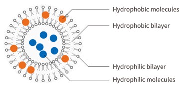
Figure 1. Liposome structure
Depending on the purpose of the research, modifications can be made to the components of liposomes (such as different phospholipid and cholesterol content), and their surface charge (generated by the charge of the constituent phospholipids), etc. This allows for the preparation of long-circulating liposomes (such as PEG-modified) for extended circulation time, cationic liposomes (modified to carry a positive charge) as gene drug carriers, environmentally sensitive liposomes (changes of in vivo pH, enzymes, or physical conditions such as light or heat effects) that respond to specific sites for drug release, and immunoliposomes that improve targeting (for example, by connecting antibodies) [1,2]. Based on the above characteristics, H. Nsairat et al. [1] summarized the clinical applications of liposomal drugs products on the market. The primary application is in anti-tumor treatments, as detailed in Table 1.
Liposomal drugs, as carrier nanomedicines, compared to conventional dosage forms, have special nanoparticle size, phospholipid bilayer structure, and special surface modifications, which cause changes in the physicochemical properties and biological behaviors. The in vivo absorption characteristics of conventional dosage forms are mainly reflected by the determination of the concentration of free drugs in the systemic circulation, while the difference between liposomal drugs products and conventional dosage forms lies in the existence of their functional unit "drug particles". After entering the body, drugs exist in the form of free (or non-liposomal) drugs substance (the drugs released from liposomes mainly exist in two forms: free drug and plasma protein binding drug in the body) and liposome-associated drugs (the drugs contained within liposomes), as shown in Figure 2. Therefore, it is necessary to determine the concentration of non-liposomal and liposome-associated drugs in blood or plasma respectively when conducting pharmacokinetics (PK) studies [3].
Table 1. Clinically Used Liposomes Grouped by Therapeutic Usage
Usage | Trade Name | Active Ingredient(s) | Liposome Platform | Manufacturer | Year Approved |
Anti-Cancer | Doxil® | Doxorubicin | HSPC:Cholesterol:PEG 2000-DSPE (56:38:5) | Sequus Pharmaceuticals | 1995 |
DaunoXome® | Daunorubicin | DSPC:Cholesterol (2:1) | NeXstar Pharmaceuticals | 1996 | |
Depocyt® | Cytarabine | DepoFoamTM | SkyPharma Inc. | 1999 | |
Myocet® | Doxorubicin | Cholesterol:EPC (45:55) | Elan Pharmaceuticals | 2000 | |
Mepact® | Mefamurtide | DOPS:POPC (3:7) Multilamellar liposome | Takeda Pharmaceutical Limited | 2004 | |
Lipodox® | Doxorubicin | DSPC:Cholesterol:PEG 2000-DSPE (56:39:5) | Sun Pharma | 2012 | |
Marqibo® | Vincristine | SM:Cholesterol (60:40) | Talon Therapeutics | 2012 | |
OnivydeTM | Irinotecan | DSPC:Cholesterol:MPEG-2000-DSPE (3:2:0.015) | Merrimack Pharmaceuticals | 2015 | |
Lipusu® | Paclitaxel | NA | Luye Pharma Group | 2006 | |
Vyxeos® | Cytarabine:Daunorubicin | DSPC:DSPG:Cholesterol (7:2:1) | Jazz Pharmaceuticals | 2017 | |
Anti-Fungal | Ambisome® | Amphotericin B | HSPC:Cholesterol:DSPG (2:1:0.8) | Astellas Pharma | 1997 |
Fungisome® | Amphotericin B | PC:Cholesterol (7:3) | Lifecare Innovations | 2003 | |
Photodynamic therapy | Visudyne® | Verteporphin | Verteporphin:DMPC&EPG (1:8) | Novartis AG | 2000 |
Analgesic | DepoDuTM | Morphine sulfate | DepoFoamTM | SkyPharma | 2004 |
Exparel® | Bupivacaine | DepoFoamTM | Pacira pharmaceuticals | 2011 |
Due to the specific clinical advantages of liposomal formulations and their unique PK characteristics, in vivo drug research on this formulation has become increasingly hot in recent years. As regulatory agencies become more mature regarding the non-clinical PK of nanoparticle technology, it is necessary to establish a reproducible, robust method that can accurately determine free and liposomal-entrapped drugs in biological samples.
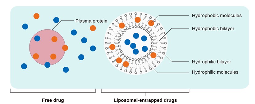
Figure 2. The Existence Form of Liposomal Drugs in Plasma
During bioanalytical method development, the separation of free and liposomal-entrapped drugs, as well as ensuring the stability of liposomes in the biological matrix during sample storage and pretreatment, present unique challenges compared to conventional dosage forms. The preparation of calibration standards and quality control samples (QCs), along with evaluations of matrix effect, selectivity, recovery, stability, and even centrifugal speed, require different considerations from those of conventional dosage forms. Based on regulatory understanding and existing project experience, this section will introduce the key points in the detection process of biological samples of this type of drug.
Bioanalytical Method Development Strategies for Liposomal Drugs
Compared with conventional dosage forms, the existence morphology of liposomal drugs in the body is complicated. Therefore, the following sections will introduce the critical points that need additional attention during the method development for bioanalysis.
Separation Method
For the determination of free and liposomal-entrapped drugs in vivo, whether using a direct method (determination of free and liposomal-entrapped drugs) or an indirect method (determination of total and free drugs), accuracy depends on sample processing. Both approaches must ensure that the state of different morphological components, such as loaded particles and free drugs, does not change. Several methods have been proposed to isolate free (or non-liposomal) and liposomal drugs, such as equilibrium dialysis, ultracentrifugation, ultrafiltration, and solid-phase extraction (SPE) [3]. Appropriate methods should be selected based on specific experimental objectives. We briefly introduce these types of methods, as detailed in Table 2.
Table 2. Separation Method Selection
Separation Method | Separation Principle | Separation State | Application |
Equilibrium Dialysis | On both sides of the semi-permeable membrane, only small molecules are allowed to pass through, while large molecules cannot. | F-F permeates the membrane, and F-P is retained in the L section. | Compounds that do not adsorb to the semipermeable membrane, and the liposomes stable in this buffer solution |
Ultracentrifugation | Different intensities of centrifugal force cause substances of different masses to separate in stages. | F-F, F-P, and L can all be separated. | Suitable for liposome formulations of hydrophilic compounds |
Ultrafiltration | Under the action of centrifugal force, small molecule drugs can pass through the ultrafiltration membrane, while large molecules are retained. | F-F permeates the membrane, and F-P is retained in the L section | Suitable for hydrophilic compounds with a lower protein binding rate in biomatrix |
Solid-phase Extraction | The electrostatic or hydrophobic interaction forces between the packing material and the compounds retain the free compounds on the packing material, while the loaded drugs pass directly through without being retained. | F and L can be separated. | Suitable for these compounds that can be adsorbed by the stationary phase of SPE for biomatrix. |
* F: Free drug; F-F: The free drug that is not bound to plasma proteins; F-P: The free drug that is bound to plasma proteins; L: Liposomal-entrapped
The equilibrium dialysis method is time-consuming. Furthermore, for drugs with a high protein binding rate, the concentration of the free form in the free state is low, which requires a higher sensitivity for the detection method. The high centrifugal force of the ultracentrifugation method may lead to the agglomeration of particles, the destruction of the bilayer structure of liposomes, and drug leakage. Solid-phase extraction requires that the test substance can be adsorbed by the solid-phase packing, and that the components on the surface of the liposome bilayer do not interact with the solid phase packing, and flow out smoothly through the gaps between the packing particles. Ultrafiltration is suitable for hydrophilic compounds with a low protein binding rate and complements the solid-phase extraction method. Based on the above methods, it is possible to choose according to actual work requirements. Solid-phase extraction and ultrafiltration are commonly used separation methods for liposomal drug DMPK bioanalysis (the operation steps are illustrated in Figures 3 and 4).
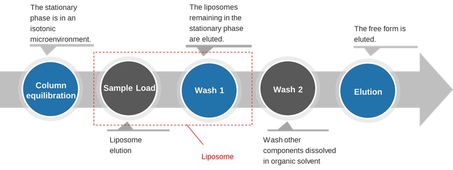
Figure 3. General Process of SPE
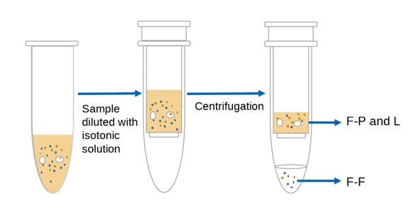
Figure 4. General Process of Ultrafiltration
Preparation of Calibration Standard Samples
The "Technical Guidance for Non-clinical Pharmacokinetic Studies of Nanomedicine (Draft)" of China points out that, when establishing the in vivo analysis method for nanomedicine carriers, it is recommended to prepare the calibration standards and quality control samples by simulating the actual in vivo state of loaded particles, free drugs, liposomal-entrapped drugs, and carrier materials.
For total drug determination: To preliminarily understand the concentration differences between free and liposomal-entrapped drugs at different time points of in vivo samples according to the results of the preliminary test, it is recommended to simulate the in vivo state of free and liposomal-entrapped drugs after administration and prepare calibration samples according to a certain proportion.
For free drug determination: Considering the low in vivo concentration of free drug (generally < 20% of the total drug in the concentration) and the possible influence of liposome encapsulation efficiency, it is generally not recommended to prepare the calibration sample by mixing the liposome and free drug.
When the free drug is used to prepare the calibration standards, due to the presence of phospholipids and charges on the surface of coexisting liposomes, non-specific binding may occur with the drug, and it needs to be considered that the coexisting liposomes may cause the free drug to be eluted from SPE.
For liposomal-entrapped drug determination: it is required that different vial liposome formulations of the same lot have consistent encapsulation efficiency and good stability in the solution state. Liposomes can be directly used to prepare calibration standards into the matrix. Since it is necessary to simulate the in vivo state, and their concentration is generally several times or even tens of times of the free drug, then it is necessary to investigate the stability of liposomes during the pre-treatment process, as well as the impact on the concentration of the free drug in case of leakage.
The Additional Focus of Validation Items
Referring to the validation items involved in the implementation recommendation of ICH M10: Bioanalytical Method Validation and Study Sample Analysis of International Council for Harmonisation of Technical Requirements for Pharmaceuticals for Human Use, and according to the actual work requirements, we summarized the areas requiring additional attention in Table 3.
Table 3. Additional Validation Considerations in Bioanalytical Method Development[4]
Validation Items | Conventional Dosage Forms | Liposomal Drugs | ||
Total Drug | Free Drug | Liposome-Associated Drug | ||
Preparation of Calibration Standards and QCs | Matrix and free drug | Matrix, free drug, and liposome-associated drug | Matrix and free drug | Matrix and liposome-associated drug |
Matrix effect | Six sources of blank matrix and one source of hemolytic matrix | Six sources of blank matrix and one source of hemolytic matrix | Extra attention should be paid to the impacts brought by the high concentration of blank liposome formulation. | Six sources of blank matrix and one source of Hemolytic matrix |
Recovery | Normal preparation | Normal preparation | Extra attention should be paid to the impacts brought by the high concentration of blank liposome formulation. | Normal preparation |
Stability | Normal preparation | Normal preparation | Extra attention should be paid to the impacts brought by liposomes after storage and freeze-thawing. | Normal preparation |
Selectivity | Six sources of blank matrix | Extra attention should be paid to the impacts brought by the high concentration of blank liposome formulation | ||
Since liposomal drugs contain both the test article and other formulation components in the sample after administration, it is necessary to consider the impact of the formulation components when preparing simulated biological samples for validation. A plasma matrix containing blank liposomes (it is recommended to choose the amount of blank formulation at the total drug ULOQ concentration, or the maximum concentration of blank liposome formulation from which the free drug has been eluted) can be used for the investigation of selectivity and matrix effect.
The special components of liposomal formulations lead to the liposome surface easily adsorbing hydrophobic compounds. When solid-phase extraction is used for separation, liposomes may also compete with the stationary phase of the column to adsorb hydrophobic compounds, so that part of the free drug enters the loading buffer along with the liposomes, resulting in inconsistent recovery of free drug at low and high concentrations. Therefore, the recovery of free drug in the presence of blank liposomes needs to be investigated during method development, meanwhile, the maximum blank liposome formulation concentration that causes free drug to be eluted should also be investigated. Therefore, during the detection of plasma samples of free drugs after administration, the samples containing high-concentration liposomes (if the concentration of liposome samples are higher than the maximum concentration) should be diluted to the appropriate concentration range for detection.
In terms of stability evaluations, it is necessary not only to consider the stability of free drug at different temperatures in the matrix, but also to pay attention to whether the introduction of additives (stabilizers, anticoagulants, etc.) will cause the destruction of liposome structure. Therefore, when investigating the stability conditions of the free drug, attention should also be paid to the stability of the liposomes under the same conditions, to prevent the occurrence of "false-positive" conclusions about the stability of the free drug caused by minor leakage from the liposomes.
Centrifuge Speed
For nanoliposomes freeze-dried powder, typically forms a suspension after dissolution. For the blood samples collected after administration, stratification of the nanoliposomes may occur after a period of time. Then during the preparation of plasma samples by centrifugation, there may be uneven distribution of liposomes in the plasma. Therefore, the effect of centrifugal force on liposome delamination and leakage needs to be investigated.
The difference in the peak areas of liposomes and free drugs in plasma at different centrifugal forces can be used to determine whether the homogeneity of liposomes is affected, so that the appropriate centrifugal force can be selected for the preparation of plasma samples.
In addition, the drug may leak from the liposome during vortex mixing, so it is necessary to gently mix the sample and working solution as low as possible during bio-sample preparation.
Summary
Liposomal drug product components are complex, and the physicochemical properties of different surface modifications vary significantly in in vivo studies. Thus, the considerations in the process of bioanalytical method development still need to be "drug-specific". However, the core principle remains finding an appropriate separation method for the separation of free and liposomal-entrapped drugs, and it is necessary to ensure that the liposomes remain in a stable state from collection to treatment and do not affect the detection of free drugs during DMPK bioanalysis.
Authors:Peiyun An, Jinlian Lu, Lili Xing
Talk to a WuXi AppTec expert today to get the support you need to achieve your drug development goals.
Committed to accelerating drug discovery and development, we offer a full range of discovery screening, preclinical development, clinical drug metabolism, and pharmacokinetic (DMPK) platforms and services. With research facilities in the United States (New Jersey) and China (Shanghai, Suzhou, Nanjing, and Nantong), 1,300+ scientists, and over fifteen years of experience in Investigational New Drug (IND) application, our DMPK team at WuXi AppTec are serving 1,700+ global clients, and have successfully supported 2,100+ IND applications.
Reference
[1] Hamdi Nsairat, Dima Khater, Usama Sayed, Fadwa Odeh, Abeer Al Bawab, Walhan Alshaer, Liposomes: structure, composition, types, and clinical applications, Heliyon, 8 (2022) e09394.
[2] Pran Kishore Deb, Omar Al-Attraqchi, Balakumar Chandrasekaran, Anant Paradkar and Rakesh K. Tekade, Protein/Peptide Drug Delivery Systems: Practical Considerations in Pharmaceutical Product Development, Basic Fundamentals of Drug Delivery, 2019: 651-684.
[3] Center for Drug Evaluation, NMPA, “Guiding Principles for Nonclinical Pharmacokinetic Studies of Nanodrugs (Trial)”, Aug,27.2021
[4] Liang Shen, editor. Drug Metabolism and Pharmacokinetics: Frontiers, Strategies, and Applications. Wiley. 2025. http://doi.org/10.1002/9781394300150.


Related Services and Platforms


-
 DMPK BioanalysisLearn More
-
Novel Drug Modalities DMPK Enabling PlatformsLearn More
-
Novel Drug Modalities BioanalysisLearn More
-
Small Molecules BioanalysisLearn More
-
Bioanalytical Instrument PlatformLearn More
-
PROTAC DMPK ServicesLearn More
-
ADC DMPK ServicesLearn More
-
Antibody-Oligonucleotide Conjugate (AOC) DMPK ServicesLearn More
-
Oligo DMPK ServicesLearn More
-
PDC DMPK ServicesLearn More
-
Peptide DMPK ServicesLearn More
-
mRNA DMPK ServicesLearn More
-
Covalent Drugs DMPK ServicesLearn More

Stay Connected
Keep up with the latest news and insights.

