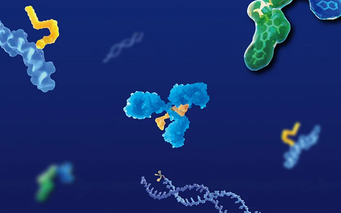
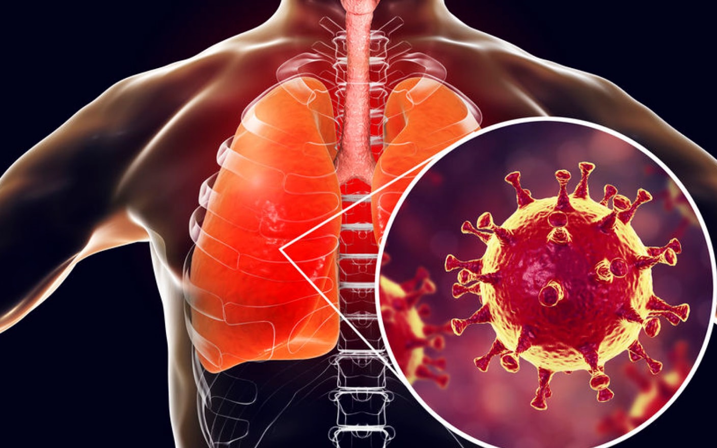
Antibody-drug conjugates (ADCs) are like precision-guided missiles, with payloads as the explosive warhead. The success of ADC therapy depends on how many payloads are delivered to the target tissues and successfully released there. Toxicities observed with ADCs may be associated with the exposure of payloads in non-target tissues.
Quantitative Whole-body Autoradiography (QWBA) is a specialized technique for analyzing the distribution of radiolabeled compounds in animal tissues. It is routinely used to determine the tissue distribution of new drug candidates in toxicological species and to assess the maximum radioactive dose for human AME research. In recent years, QWBA has been frequently used to obtain the target tissue specificity of ADC drugs.
Case Study
Here shows a case study to illustrate how to use the QWBA technique to investigate the tissue distribution of ADC in tumor-bearing nude rats. The structure of [3H] DM1-LNL897 is shown in Figure 1.
![Structure of [3H] DM1-LNL897 and parameters of QWBA study](https://wuxiapptec-dmpkcatalog-prod.oss-cn-shanghai.aliyuncs.com//upload/image/20230816/483987565947356.png)
Figure 1. Structure of [3H] DM1-LNL897 and parameters of QWBA study
In this case, the ADC was radiolabeled with tritium in the payload. After intravenous administration in tumor-bearing nude rats, QWBA was used to analyze the tissue distribution of payloads at different time points.1 Representative QWBA images at 4 time points were selected to illustrate the tissue distribution of radioactivity-related components in tumor-bearing nude rats after administration of the ADC (Figure 2).
![Tissue distribution of radioactivity in tumor-bearing nude rats at 1 h and 24 h post-dose after [3H] DM1-LNL897 administration](https://wuxiapptec-dmpkcatalog-prod.oss-cn-shanghai.aliyuncs.com//upload/image/20230816/544395454767748.png)
Figure 2. Tissue distribution of radioactivity in tumor-bearing nude rats at 1 h and 24 h post-dose after [3H] DM1-LNL897 administration.1
At 1h post-dose, radioactivity was mainly concentrated in the blood-rich tissues, such as blood, heart, and liver, radioactivity also started to enter tumor tissues. At 24h post-dose, the concentration of radioactivity in the tumor increased significantly, reaching a peak that far exceeded other tissues. There was little evidence of radioactivity in the skin, but essentially no radioactivity entered the brain or muscle tissues.
At 72h post-dose, the image on the left showed that the payloads, including intact ADC and released payloads, were mainly concentrated in the tumor as the target tissue. Besides the tumor, there was no accumulation or tissue retention of radioactivity in any of the tissues analyzed, demonstrating the good selectivity of the ADC. The autoradiograph on the right showed the radioactivity exposure of non-target tissues, demonstrating that the concentration of radioactivity had decreased continuously. At 168 h post-dose, the tumor tissues still contained payload-related radioactivity (Figure 3).
![Tissue distribution of radioactivity in tumor-bearing rats at 72 h and 168 h after [3H] DM1-LNL897 administration](https://wuxiapptec-dmpkcatalog-prod.oss-cn-shanghai.aliyuncs.com//upload/image/20230816/558659506379105.png)
Figure 3 Tissue distribution of radioactivity in tumor-bearing rats at 72 h and 168 h after [3H] DM1-LNL897 administration.1
The use of QWBA to study the tissue distribution of payloads in tumor-bearing nude rodents provides an effective research approach for preclinical assessments of the efficacy in target tissues and the organ toxicity of ADC.
DMPK’s QWBA Introduction
After animals were given a radiolabeled test article, QWBA was applied to study the distribution of total radioactivity throughout their bodies to obtain complete and detailed tissue distribution results and to calculate the maximum radioactive dosage that can be administered to humans based on exposure and half-life in each tissue.
Whole blood was collected from one animal at each of the specific time points after dosing, and carcasses were frozen in liquid nitrogen. Sagittal sections of each animal were created using a whole-body cryomicrotome (Leica CM3600 XP Microsystems). The representative sections were exposed to close contact with phosphor screens. Quantitative whole-body autoradiography was performed using the phosphor image technique using Typhoon GRB scanner and AIDA Image Analyzer.
WuXi AppTec DMPK’s radioactivity platform provides a variety of animal models, including CD-1 mice, C57 mice, SD rats, LE rats, cynomolgus monkeys, etc. for QWBA research.
Click here to learn more about the strategies for ADC, or talk to a WuXi AppTec expert today to get the support you need to achieve your drug development goals.
Authors: Xue Yu, Wei Lei
Committed to accelerating drug discovery and development, we offer a full range of discovery screening, preclinical development, clinical drug metabolism, and pharmacokinetic (DMPK) platforms and services. With research facilities in the United States (New Jersey) and China (Shanghai, Suzhou, Nanjing, and Nantong), 1,000+ scientists, and over fifteen years of experience in Investigational New Drug (IND) application, our DMPK team at WuXi AppTec are serving 1,500+ global clients, and have successfully supported 1,200+ IND applications.
Reference
1.Markus Walles, Bettina Rudolph, Thierry Wolf, etc. New Insights in Tissue Distribution, Metabolism and Excretion of [3H]-labeled Antibody Maytansinoid Conjugates in Female Tumor Bearing Nude Rats. ASPET Journals, 2016
Related Services and Platforms


-

 Radiolabeled In Vivo ADME StudyLearn More
 -

 Novel Drug Modalities DMPK Enabling PlatformsLearn More
 -

 Radiolabeled MetID (Metabolite Profiling and Identification)Learn More
 -

 Radiolabeled Non-Clinical In Vivo ADME StudyLearn More
 -

 Quantitative Whole-body Autoradiography (QWBA)Learn More
 -

 Human Radiolabeled Mass Balance StudyLearn More
 -

 Radiolabeled Compound SynthesisLearn More
Stay Connected
Keep up with the latest news and insights.

