In drug development, assessing cytochrome P450 (CYP) enzyme inhibition is crucial to predict potential drug-drug interactions (DDIs). Traditionally, liver microsomes have been the gold standard for CYP inhibition screening due to their simplicity and cost-effectiveness. However, as our understanding of drug metabolism evolves, there is a growing need to incorporate more physiologically relevant models, such as hepatocytes, for improving translational accuracy.
This blog discusses the strengths and limitations of both microsomal and hepatocyte systems, reasons for potentially weaker inhibition values from the assay with hepatocyte, and explores how to optimize DDI predictions with both microsomal and hepatocyte-based assays. It also introduces a new hepatocyte-based CYP inhibition screening service, designed to complement existing microsomal assays and enhance clinical risk assessment (Table 1).
Table 1. Study design of CYP inhibition studies using human hepatocytes, which enables assessment of risk potential, supports decision-making, and informs definitive design.
Study Design | Standard Study Report Includes: |
|
|
Microsomes vs Hepatocytes in CYP Inhibition Studies
Microsomes: The Gold Standard for Early Screening
Liver microsomes, as subcellular fractions enriched with CYP enzymes, become highly efficient for high-throughput screening when supplemented with essential cofactor systems such as the NADPH regeneration system. They are widely used to assess:
Direct inhibition (reversible binding of inhibitors to CYP enzymes)
Time-dependent inhibition (TDI) (mechanism-based inactivation requiring metabolic activation)
Key advantages of microsomes include:
High enzyme concentration→ Enhanced sensitivity for detecting inhibitors
Minimal interference from cellular uptake/efflux transporters
Cost-effective and scalable for early-stage screening
However, the absence of intact cellular architecture in microsomes leads to several limitations in CYP inhibition assessment:
Membrane permeability effects
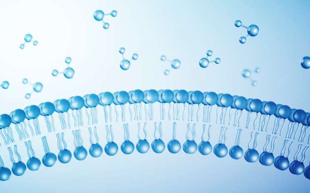
The lack of complete cell membranes may artificially enhance the apparent potency of poorly permeable compounds, resulting in lower (more potent) IC50 values than would be observed in cellular systems.
Transport and metabolic compensation
Microsomal systems cannot account for:
Transporter-mediated uptake/efflux (e.g., OATP/P-gp)
Competing non-CYP metabolic pathways (e.g., UGT, SULT). Consequently, inhibition data may diverge significantly from hepatocyte results - potentially overestimating or underestimating true inhibitory potential.
Hepatocytes: A More Physiologically Relevant Model
Primary hepatocytes retain intact cellular architecture, including:
CYP enzymes in their native environment
Transporters (e.g., OATPs) that may influence drug exposure
Cellular defense mechanisms (e.g., glutathione conjugation)
Our new hepatocyte-based CYP inhibition service provides:
More clinically translatable inhibition values (closer to in vivo effects)
Better prediction of intracellular drug concentrations (accounting for uptake/efflux)
Improved assessment of metabolite-mediated TDI (due to full metabolic competence)
Key Differences in CYP Inhibition Results: Why Hepatocyte Data May Appear Weaker
A. Direct CYP Inhibition (Reversible Inhibition)
In hepatocytes, observed inhibition is often weaker than in microsomes due to:
Nonspecific binding (reducing free drug concentration)
Competing elimination pathways (e.g., Phase II metabolism, biliary excretion)
Intracellular sequestration (e.g., lysosomal trapping)
Example: Ketoconazole, showing an IC50 of 0.00745 μM in microsomes (Figure 1), may yield an IC50 of 0.126 μM in hepatocytes (Figure 2) due to reduced free intracellular concentration.

Figure 1. Direct inhibition of CYP3A-Midazolam in microsomes

Figure 2. Direct inhibition of CYP3A-Midazolam in hepatocytes
B. Time-Dependent Inhibition (TDI)
For mechanism-based inhibitors, hepatocytes may show:
Slower inactivation kinetics (due to lower free inhibitor concentration)
Possible detoxification (e.g., conjugation of reactive metabolites)
Example: The IC50 Shift value of Azamulin is both greater than 1.50 in microsomes (Figure 3) and in hepatocytes (Figure 4); however, the kinact/KI value is much larger in the microsomes than in hepatocytes. (Table 2)

Figure 3. Time-dependent inhibition of CYP3A-Midazolam in microsomes

Figure 4. Time-dependent inhibition of CYP3A-Midazolam in hepatocytes
Table 2. The Kinact/KI value of Azamulin towards CYP3A was generated in two different test systems
Test System | CYP Isoforms | Inhibitor | kinact/KI(µM-1min-1) |
HLM | CYP3A | Azamulin | 1.18 |
Hepatocytes | CYP3A | Azamulin | 0.159 |
Conclusion: A Two-Tiered Approach for Optimal CYP Inhibition Assessment
To maximize efficiency and accuracy, we recommend:
Step 1: Microsomal Screening – Identify potent inhibitors quickly and cost-effectively.
Step 2: Hepatocyte Confirmation – Refine inhibition estimates for clinically relevant risk assessment.
By integrating both models, we can reduce attrition in drug development and improve the accuracy of DDI predictions.
Are you interested in learning more about our hepatocyte-based CYP inhibition screening service? Please contact us to discuss how we can support your drug development program.
Talk to a WuXi AppTec expert today to get the support you need to achieve your drug development goals.
Committed to accelerating drug discovery and development, we offer a full range of discovery screening, preclinical development, clinical drug metabolism, and pharmacokinetic (DMPK) platforms and services. With research facilities in the United States (New Jersey) and China (Shanghai, Suzhou, Nanjing, and Nantong), 1,000+ scientists, and over fifteen years of experience in Investigational New Drug (IND) application, our DMPK team at WuXi AppTec are serving 1,600+ global clients, and have successfully supported 1,700+ IND applications.
Reference
[1] Obach, R. S., Walsky, R. L., & Venkatakrishnan, K. (2007). Drug Metabolism and Disposition. 35(2), 246-252.
[2] Wienkers, L. C., & Heath, T. G. (2005). Nature Reviews Drug Discovery. 4(10), 825-833.
[3] Fahmi, O. A., Hurst, S., Plowchalk, D., et al. (2009). Drug Metabolism and Disposition. 37(8), 1658-1666.
[4] Brown, Hayley S. et al. Drug Metabolism and Disposition, Volume 38, Issue 12, 2139 - 2146
Stay Connected
Keep up with the latest news and insights.


