

Liposome Drug Delivery: Classification, Composition, and Formulation Considerations
-

Articles
-

Jan 16, 2026
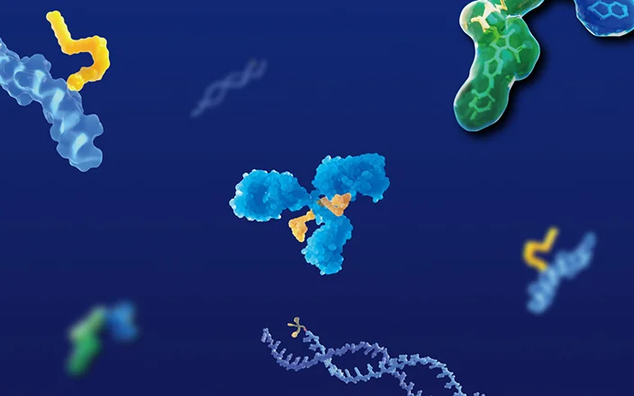
Liposome drug delivery represents a versatile and effective platform for transporting therapeutic agents. Its adaptability has led to widespread adoption in critical fields like oncology and vaccinology (Figure 1). The rapid deployment of LNP-mRNA vaccines for COVID-19, such as those from Pfizer-BioNTech and Moderna, underscores the pivotal role of liposome drug delivery in achieving timely clinical and commercial success, propelling the field forward.
Surface engineering enables these systems to be fitted with precise targeting moieties, allowing for selective binding to cellular receptors. A key advantage of liposome drug delivery is its capacity to co-encapsulate both water-soluble and lipid-soluble actives, facilitating combination therapies. This review outlines the makeup and types of liposomal systems, highlights their successful preclinical application, and discusses central PK hurdles and corresponding solutions.

Figure 1. Diverse therapeutic and research applications enabled by liposome drug delivery [1,2]
Liposome Drug Delivery: Definition and Application
Among nanoscale carrier platforms, lipid-based systems—notably liposomes and lipid nanoparticles (LNPs)—stand out as particularly successful. LNPs represent an evolution of classic liposome design, sharing structural similarities but incorporating a multilayered core formed via electrostatic interaction between cationic lipids and nucleic acids. This advancement has made them indispensable for nucleic acid transport, cementing their status as a focal point in modern drug delivery research.

Figure 2. Classification of lipid-based nanocarrier systems: 1. Liposomes 2. Lipid nanoparticles (LNPs) 3. Solid lipid nanoparticles (SLNs) 4. Hybrid lipid nanoparticles [2]
Structurally, liposomes are spherical vesicles with one or more phospholipid bilayers enclosing an aqueous interior. Their success in the pharmaceutical sector is evident, particularly in oncology and vaccine development. Over two dozen liposomal medicines have gained market approval since the mid-1990s. Figure 3 illustrates how pioneering products have expanded the reach of liposome drug delivery into diverse therapeutic areas, including antimicrobial therapy, ophthalmology, analgesia, and immunology.

Figure 3. Key commercialized products utilizing liposome drug delivery technology [3]
Classification and Composition of Liposome Drug Delivery Systems
Liposomes can generally be classified into conventional liposomes and specialized liposomes. The specialized liposomes, such as pH-sensitive liposomes, immunoliposomes, and cationic liposomes, are optimized to meet specific application needs.
Table 1. Classification of liposome drug delivery systems and their profiles
Liposome Type | Composition and Major Features | Applications |
Conventional liposomes | Neutral or negatively charged phospholipids, cholesterol | Foundational liposomal delivery for various drugs and diagnostic agents |
Long-circulating liposomes | Surface-modified liposomes with inert, highly biocompatible polymers (e.g., PEGylation) | Enhanced bioavailability, suitable for sustained drug release therapies |
pH-sensitive liposomes | Structural changes in specific pH environments to release encapsulated drugs | Targeted delivery to tumor tissues (usually lower pH) or specific physiological environments (e.g., intestines) |
Cationic liposomes | Composed of cationic lipids suitable fo loading negatively charged macromolecules (e.g., DNA, RNA, oligonucleotides) | Used as gene delivery vectors to enhance gene transfection efficiency |
Targeted liposomes | Surface-modified with specific ligands (e.g., antibodies, peptides, small molecules) to target specific cells or tissues | In cancer treatment, targeted liposomes can selectively deliver drugs to tumor cells, reducing the impact on healthy cells. |
The key components of liposomes mainly include phospholipids and cholesterol. Phospholipids, the primary components of biological cell membranes, exhibit good biocompatibility and degradability. Moreover, they are endogenous substances in vivo, typically non-toxic and non-immunogenic. The final formulation generally uses only 1 to 2 types of phospholipids to simplify the characterization and scale-up production of liposomal delivery-based therapeutic products.
(1) Phospholipids
As the core components of liposomes, the structure and properties of phospholipids directly influence the function and applications of liposomes. The structures of phospholipids have common features: hydrophilic polar head groups and hydrophobic tails. Hydrophobic fatty acid chains self-assemble in aqueous phases to form bilayers, constituting the basic structure of liposomes. Based on different phospholipid headgroups, phospholipids can be classified into positively charged phospholipids (lecithin (PC) and phosphatidylethanolamine (PE)), negatively charged phospholipids (phosphatidylglycerol and phosphatidylserine), and neutral phospholipids. The surface charge of liposomes, determined by the charge properties of their constituent phospholipids, influences their behavior in vivo studies, including drug liposomal delivery efficiency, cellular uptake, and interactions with other biological components.

Figure 4. Phospholipid structure[4]
(2) Helper Lipids and mRNA-LNPs
Helper lipids are non-primary phospholipid components used to enhance liposome performance, stability, and liposomal delivery capacity. Helper lipids typically work in conjunction with primary phospholipids to improve liposome characteristics. Helper lipids include PEGylated lipids and cholesterol, which have unique functional properties (Table 2).
Table 2. Functional roles of lipid components in liposome drug delivery
Lipid composition | Functional features | |
phospholipid | Anionic lipids | Electrostatic interactions with cationic lipids or drugs enhance liposome stability and drug loading capacity. |
Cationic lipids | Electrostatic interactions with negatively charged nucleic acids efficiently encapsulate nucleic acids. | |
Ionizable cationic lipids | Change charge depending on environmental pH, avoiding blood clearance and immune system stimulation. | |
Secondary lipids | Cholesterol | Regulate membrane fluidity and rigidity, increasing liposome stability |
PEGylated lipids | Reduce liposome binding to plasma proteins and rapid elimination by the reticuloendothelial system, prolonging circulation time; enhancing bioavailability. | |
LNPs are specialized liposomes for mRNA delivery, widely used in vaccine development and gene therapy. In LNPs, helper lipids play a crucial role, significantly improving LNP stability, efficacy, and biocompatibility. As shown in Figure 5, three marketed mRNA-LNPs primarily consist of ionizable cationic lipids, phospholipids, and helper lipids (cholesterol and PEGylated lipids).

Figure 5. Composition of three approved mRNA-LNPs [5]
Challenges and Formulation Optimization Strategies of Liposome Drug Delivery Systems
Liposomes hold broad application potential in preclinical formulation screening but face several challenges. The selection of lipid components is a critical step in preclinical liposome development, directly influencing liposome physicochemical properties, drug loading capacity, biocompatibility, stability, and therapeutic efficacy. During the early stages of liposome development, thorough safety evaluations of selected lipid components (such as anionic phospholipids, cationic lipids, helper lipids, etc.) are necessary to ensure that these components do not cause toxicity at anticipated usage levels.
Screening solvents for poorly soluble lipid components
The solubility of lipid components is primarily influenced by their chemical structure and composition. Due to the hydrophilic head and hydrophobic tail structure of phospholipids, their solubility in aqueous phases is typically low. In animal dosing experiments, addressing lipid solubility issues and considering solvent component tolerance ranges within animals are crucial. Additionally, the administration method (e.g., intravenous injection and subcutaneous injection) must meet in vivo formulation requirements, including osmotic pressure, stability, and precipitation post-intravenous administration.
Table 3. Common lipid components and solvents for liposome drug delivery systems
Common Lipid Components | Common Solvents |
Anionic lipids: HSPC (hydrogenated soy phosphatidylcholine), DSPC (distearoyl phosphatidylcholine), DOPC (dioleoyl phosphatidylcholine), DPPG (dipalmitoyl phosphatidylglycerol), DSPG (distearoyl phosphatidylglycerol), DOPE (dioleoyl phosphatidylethanolamine) | Solvents: ethanol EtOH, dimethyl sulfoxide DMSO, polyethylene glycol 400 PEG400, propylene glycol PG |
Ionizable cationic lipids: SM-102 (heptadecanoic acid ester of 8-[2-hydroxyethyl][6-O-6-(undecyl)hexyl]amino]octanoic acid), ALC-0315 ((4-hydroxybutyl)azanediyl bis(hexane-6,1-diyl bis(2-hexyldecanoate)) | Surfactants: Tween 80, polyethylene glycol (15)-hydroxystearate solutol HS15 |
Cholesterol: CHO | Aqueous phase: saline, sterile water |
PEGylated lipids: DSPE-MPEG2000 (distearoyl phosphatidylethanolamine-PEG2000), DMG-PEG2000 (1,2-dimyristoyl-rac-glycerol-3-methoxy PEG2000) | Others: cyclodextrins, cellulose, buffers, oils |
Storage stability regulation of liposomes/LNP formulations
Ensuring liposome physical and chemical stability before dosing in in vivo pharmacokinetic (PK) studies directly affects liposome drug release and bioavailability. Using inert gas protection during storage, such as dry ice, is common, but during liposome freeze-thaw processes, CO2 dissolution in solutions can lower pH, destabilizing liposomes. Air displacement methods to remove CO2 before preparation are needed. Moreover, temperature, pH, and light exposure are key environmental factors that affect the storage stability of liposomes.
Temperature: Low-temperature storage generally improves liposome stability, but excessively low temperatures can cause liposome phase changes.
pH: Liposome stability is affected by pH, typically controlled between 7 and 8. Appropriate buffers, such as Tris buffer, help maintain stable pH environments.
Light exposure: Light exposure can degrade liposome components; hence, light protection measures or avoiding light exposure are necessary.
PK studies of liposomes/LNP formulations: Pre-administration assessment
Due to liposomes' instability, assessing liposome quality before dosing in preclinical formulation studies is crucial. Ensuring study validity and reliability involves the following steps:
Physicochemical property assessment: Before dosing, check liposome formulation appearance, ensuring no precipitation, bubbles, or discoloration. Use dynamic light scattering (DLS) and other techniques to measure liposome size distribution and zeta potential, assessing corresponding physicochemical properties to ensure liposome stability.
Drug concentration confirmation: Accurately measure drug concentration in liposomes to ensure it meets experimental design requirements.
Lipid component identification and quantification: Identify and quantify individual lipid components in liposomes to ensure composition accuracy and consistency.
Common lipid components lack significant chromophores, making traditional UV detectors ineffective. Currently, high-performance liquid chromatography with charged aerosol detection (HPLC-CAD) and LC-MS are mainly used for lipid component detection[8]. We tested four most widely used commercial lipids: hydrogenated soy phosphatidylcholine (HSPC), distearoyl phosphatidylcholine (DSPC), dioleoyl phosphatidylcholine (DOPC), and cholesterol (CHO), and developed an HPLC-CAD method to detect individual lipid components and liposomes (Figure 6).

Figure 6. Chromatograms of blank solution and liposome sample solution
Final Words
Liposome drug delivery development in preclinical settings faces multiple challenges, but by evaluating and optimizing lipid components, enhancing stability, and establishing a robust quality assessment system, liposomal delivery efficiency and success rates can be effectively improved. WuXi AppTec DMPK offers extensive expertise in formulating liposome drug delivery systems, from solvent compatibility assessment to stability assurance. Our integrated analytical platform ensures accurate quantification of both drug and lipid components, guaranteeing formulation quality before in vivo studies(Figure 7).

Figure 7. Testing platform for liposome animal formulation screening
Authors: Yifan Yang, Yu Chen, Chen Ning, Chao Zhang, Shoutao Liu
Talk to a WuXi AppTec expert today to get the support you need to achieve your drug development goals.
Committed to accelerating drug discovery and development, we offer a full range of discovery screening, preclinical development, clinical drug metabolism, and pharmacokinetic (DMPK) platforms and services. With research facilities in the United States (New Jersey) and China (Shanghai, Suzhou, Nanjing, and Nantong), 1,000+ scientists, and over fifteen years of experience in Investigational New Drug (IND) application, our DMPK team at WuXi AppTec are serving 1,600+ global clients, and have successfully supported 1,700+ IND applications.
Reference
[1] Luiz H, Oliveira Pinho J, Gaspar MM. Advancing Medicine with Lipid-Based Nanosystems Successful Case of Liposomes. Biomedicines. 2023 Feb 2; 11(2)
[2] Wu S, Lin L, Shi L, Liu S. An overview of lipid constituents in lipid nanoparticle mRNA delivery systems. Wiley Interdiscip Rev Nanomed Nanobiotechnol. 2024 Jul-Aug; 16(4):e1978.
[3] Cheng Z, Fobian SF, Gurrieri E,. Lipid-based nanosystems: the next generation of cancer immune therapy. J Hematol Oncol. 2024 Jul 19; 17(1):53.
[4] Kraft JC, Freeling JP, Wang Z, Ho RJ. Emerging research and clinical development trends of liposome and lipid nanoparticle drug delivery systems. J Pharm Sci. 2014 Jan; 103(1):29-52.
[5] Suzuki Y, Ishihara H. Difference in the lipid nanoparticle technology employed in three approved siRNA (Patisiran) and mRNA (COVID-19 vaccine) drugs. Drug Metab Pharmacokinet. 2021 Dec;41:100424.
[6] Stability characterization for pharmaceutical liposome product development with focus on regulatory considerations: An update
[7] Oude Blenke E, Örnskov E, Schöneich C, N. The Storage and In-Use Stability of mRNA Vaccines and Therapeutics: Not A Cold Case. J Pharm Sci. 2023 Feb; 112(2):386-403.
[8] Yu X, Yu C, Wu X, Cui Y,. Validation of an HPLC-CAD Method for Determination of Lipid Content in LNP-Encapsulated COVID-19 mRNA Vaccines. Vaccines (Basel). 2023 May 4; 11(5)


Related Services and Platforms


-
 In Vivo PharmacokineticsLearn More
-
Novel Drug Modalities DMPK Enabling PlatformsLearn More
-
Rodent PK StudyLearn More
-
Large Animal (Non-Rodent) PK StudyLearn More
-
Clinicopathological Testing Services for Laboratory AnimalsLearn More
-
High-Standard Animal Facilities and Animal WelfareLearn More
-
Preclinical Formulation ScreeningLearn More


Stay Connected
Keep up with the latest news and insights.

