
In Vivo Imaging for Small Animals: Deciphering the Spatiotemporal Trajectory of Drugs
-

Articles
-

Oct 24, 2025
Typically, in vivo PK studies are conducted in different species based on the requirements of various development stages, balancing research progress with experimental efficiency and cost. Traditional PK studies rely on dissection sampling and in vitro analytical detection, such as analyzing blood or tissue samples obtained by sacrificing animals at different time points. For early screening stages, this method is not only time-consuming and labor-intensive but also requires extensive experimental data to interpret changes in the drug within the body. If in vivo imaging system for small animals is employed in the early research stages, changes in the drug can be tracked in real-time in the same animal non-invasively, achieving a "transparent" view for in vivo pharmacokinetic studies. This article will systematically elaborate on the principles, advantages, and applications of in vivo imaging technology for small animal PK studies.
What is In Vivo Imaging?
In vivo animal imaging typically uses imaging methods to study biological processes qualitatively or quantitatively in living organisms (e.g., at the tissue, cellular, and molecular levels). Based on technical principles and application scenarios, it is mainly divided into the following two categories (as shown in Figure 1)[1]:
Functional imaging: Suitable for studying molecular metabolism and physiological research; includes optical imaging and radionuclide imaging (PET/SPECT).
Structural imaging: Suitable for anatomical imaging; encompasses magnetic resonance imaging (MRI), ultrasound imaging, and computed tomography (CT).
Compared to structural imaging, functional imaging better reflects the spatial and temporal distribution of cellular or gene expression, thereby "transparently" illustrating relevant biological processes, specific gene functions, and interactions within the live animal. Therefore, in vivo functional imaging technology allows observation and tracking of target cells and gene expression at the molecular and cellular levels, assesses disease progression at the whole-animal level, and aids in drug optimization and refining gene therapy strategies.

Figure 1. In vivo animal imaging technologies[2-3]
High-sensitivity bio-optical imaging systems consist of a light-tight imaging chamber, an ultra-low temperature scientific-grade CCD camera, and an intelligent analysis platform. Simultaneously, integrated experimental modules are equipped with an animal anesthesia system, a constant-temperature physiological platform, and bio-containment units, supporting full-time-domain molecular imaging research from millisecond transient luminescence to long-term metabolic observation (as shown in Figure 2). In vivo optical imaging technology primarily includes two techniques: bioluminescence imaging and fluorescence imaging (as shown in Table 1). Among them, bioluminescence imaging involves using luciferase reporter genes to label cells or DNA, causing the expressed proteases to undergo biochemical reactions with corresponding substrates, thereby generating probe light signals within the organism. This technique can capture luminescent signals without requiring an external excitation light source.
Currently, the most commonly used optical imaging enzyme is firefly luciferase (FLuc), isolated from the firefly (Photinus pyralis). It uses luciferin as a substrate and, in the presence of ATP, generates oxyluciferin, emitting green light at a wavelength of 560 nm[4]. Additionally, renilla luciferase (RLuc) and gaussia luciferase (GLuc) are two other common luciferases. They use coelenterazine as a substrate and are often combined with FLuc for dual-reporter imaging, assessing specific disease model animals from multiple angles[5].
Among these, GLuc, derived from the marine copepod Gaussia princeps, is particularly unique: it can be naturally secreted in mammalian cells and has a sensitivity approximately 1,000 times greater than FLuc or RLuc. Therefore, its levels in blood or urine can serve as biomarkers for monitoring biological events such as tumor growth, viral infection and replication, and circulating cell viability[6]. These bioluminescent proteins are often preferred over their fluorescent counterparts because mammalian tissues lack endogenous bioluminescent reactions, enabling imaging with nearly zero background[7].

Figure 2. Schematic diagram of bioluminescence imaging and fluorescence imaging
Unlike bioluminescence imaging, fluorescence imaging uses fluorescent reporter genes (such as GFP, RFP) or fluorescent dyes like Cy dyes for labeling. It utilizes the fluorescence generated by fluorescent proteins or dyes to create an internal light source and requires excitation by an external light source to capture the emitted signal (as shown in Table 1). Compared to in vivo bioluminescence imaging, fluorescence imaging is less costly and does not require injecting substrates into the animal; the fluorescent chromophores emit light signals at specific wavelengths upon excitation by their corresponding wavelength of light. As long as the fluorophore is stable, it can be excited and emit light on demand.
Furthermore, since fluorescence imaging is based on the principle of physical energy transfer, it has lower requirements for the physiological state of the experimental sample: optical imaging can be achieved whether it's a live animal, an animal carcass, or an isolated organ. However, the choice of fluorescent imaging markers is closely related to the actual imaging results. It is generally recommended to choose near-infrared markers with longer wavelengths and reduce interference from the animal's own fluorescent background. In recent years, research focus in fluorescence imaging has centered on developing fluorescent contrast agents emitting in the second near-infrared window (NIR-II, 1,000–1,700 nm) and optical instruments suitable for this spectral range, as tissue transparency is highest in this wavelength range, providing optimal image resolution at greater tissue depths (about 1 cm)[7].
In summary, bioluminescence imaging occurs only in living cells and does not involve radioactive substances, characterized by its ease of operation, intuitive results, and high sensitivity. For studies involving fluorescently labeled tissues or drugs, in vivo imaging technology can be used in the early screening stages to track the dynamic changes of drugs within the animal body in real-time and non-invasively[8].
Table 1. Comparison of bioluminescence imaging and fluorescence imaging
Method | Advantages | Disadvantages |
Bioluminescence Imaging |
|
|
Fluorescence Imaging |
|
|
Applications of In Vivo Imaging in Small Animal PK Studies
Based on the advantages of the in vivo imaging system for small animals and the practical needs in the field of rodent pharmacokinetic research, we summarize the following key application scenarios (Figure 3):

Figure 3. Application scenarios of the in vivo imaging system for small animals in pharmacokinetics
Spatiotemporal tracking of drug distribution
Conventional PK studies infer drug distribution through discrete sampling, whereas in vivo imaging enables visual tracking of differential drug accumulation in target and non-target organs throughout the entire process.
Real-time monitoring of metabolic processes and determination of PK parameters
Using dual-modal probes (e.g., simultaneously labeling the parent drug and its metabolites), the metabolic transformation of drugs in the liver and excretion pathways via the kidneys can be dynamically tracked.
Investigating drug-target interaction mechanisms
Utilizing molecular probes (such as fluorescently labeled antibodies or aptamers) that specifically bind to drug targets allows verification of drug-target binding efficiency and competitive inhibition effects.
By combining genetically engineered animal models (e.g., humanized hepatocyte mice) with in vivo imaging, human drug metabolism characteristics can be simulated more accurately, predicting potential toxicity or efficacy differences at clinical doses. For instance, using real-time imaging to track the targeting efficiency of antibody drugs in immune checkpoint humanized mice can help optimize drug structure to reduce non-specific accumulation. Furthermore, imaging data-based PK-PD models will promote the development of personalized dosing regimens, especially in the fields of nanomedicine or cell therapy. In vivo imaging can dynamically assess the stability of delivery systems and their delivery efficiency to target organs, providing direct evidence for formulation improvement. This technological trend not only aligns with the "3R principles" but also enhances drug development efficiency, offering economically viable research pathways, particularly for rare diseases or complex indications.
Case Study of In Vivo Imaging
SM-102 is a synthetic cationic lipid used for mRNA vaccine delivery, playing a significant role in mRNA vaccine development, particularly in the COVID-19 vaccine (Moderna mRNA-1273), where it serves as one of the key components enhancing the in vivo delivery efficiency of mRNA. Additionally, SM-102 is used in cancer immunotherapy, gene editing, and RNA interference therapy, providing crucial support for the development of novel nucleic acid drugs. FLuc mRNA encapsulated in SM-102 LNP (FLuc mRNA (m1Ψ)-SM102 LNP), upon delivery into cells or in vivo, enables the expression of FLuc protein. After adding the luciferin substrate, it produces yellow-green fluorescence (detection wavelength 550-570 nm).
Experimental method: After intramuscularly administering equivalent doses of FLuc mRNA (m1Ψ)-SM102 LNP to mice (n=2) (as shown in Table 2), the luciferin substrate (D-Luciferin sodium salt) was injected intravenously at different time points (2, 4, 24 h). FLuc expression levels and distribution were detected in real-time using a small animal whole-body fluorescence imaging system. Additionally, at the 4-hour time point after substrate injection, continuous imaging was performed to confirm the trend of fluorescence signal change over time.
Table 2. Experimental design
Test Article | Dose Route | Dosage | Concentration | Dose Volume | Time Points |
Fluc mRNA (m1Ψ)-SM102 LNP | IM | 15 μg/animal | 0.15 mg/mL | 0.100 mL/animal | 2h, 4h, 24h |
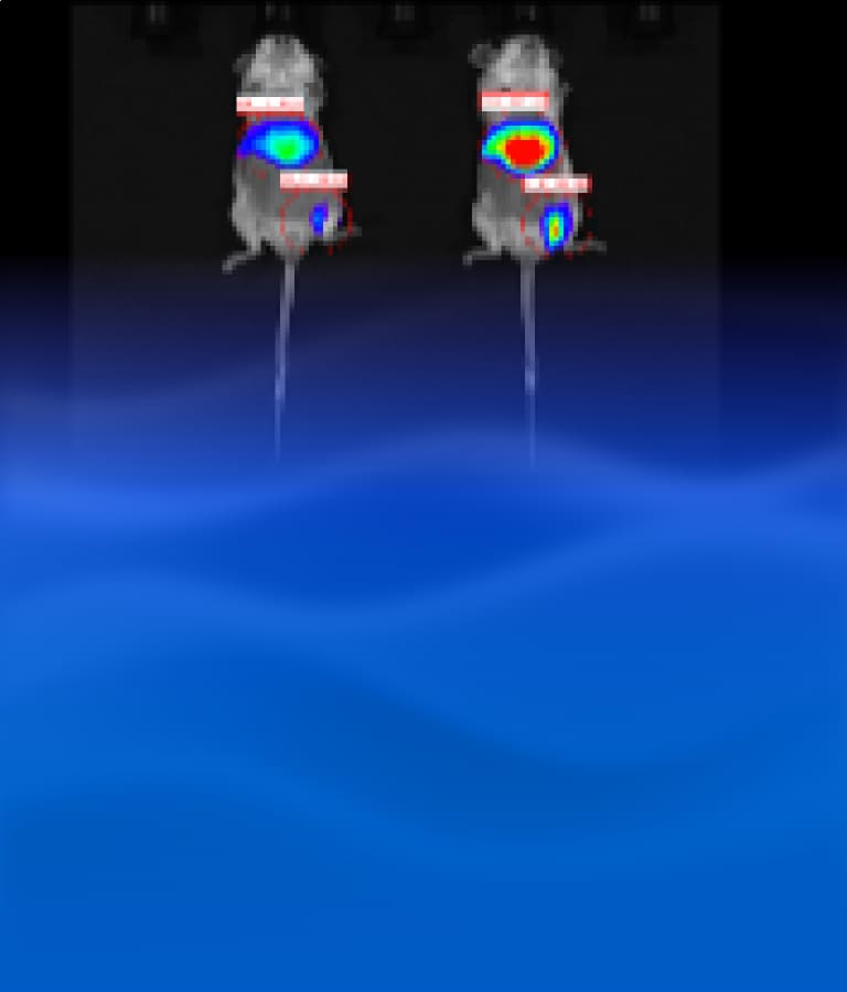
The experimental results are shown in Figures 4 and 5:
After administration, the expressed FLuc protein was primarily distributed in the liver area and the injection site.
Compared to other time points, the enrichment of Fluc mRNA in the liver and injection site was higher at 4 hours post-administration.
A relatively obvious fluorescence signal plateau was observed around 5 minutes after substrate injection. After 5 minutes, the fluorescence signal gradually declined, and within the 5-15 minutes interval, the fluorescence signal decrease rate could reach 50%.

Figure 4. Expression and distribution of FLuc in the animal body at different time points
")
Figure 5. Trend of fluorescence signal change over time 5 minutes after substrate injection (partial data)
Based on the above experimental results, in vivo imaging system for small animals can efficiently screen the in vivo distribution of drugs and reveal the dynamic trends of the drug through the numerical conversion of optical signals. However, it is noteworthy that since the fluorescence signal generated by the luciferase-catalyzed reaction decays over time, special attention must be paid to optimizing the imaging time window (i.e., the optimal time range for imaging after substrate injection) to ensure data reliability.
Concluding Remarks
With the rapid development of precision medicine and drug development technologies, the in vivo imaging system for small animals, as an important tool in preclinical pharmacokinetic research, is increasingly becoming a critical link in the innovative drug development chain. By integrating multi-modal technologies such as optical imaging (e.g., bioluminescence, fluorescence), radionuclide imaging (e.g., PET/SPECT), Magnetic Resonance Imaging (MRI), and Computed Tomography (CT), in vivo imaging systems enable non-invasive, real-time, and visual tracking of the dynamic behavior of drugs within live animals, providing multi-dimensional data support for PK and PD studies. In the future, technological breakthroughs and deeper applications in this field will continue to enhance the efficiency of drug development and promote the optimization of personalized treatment strategies. WuXi AppTec DMPK possesses extensive experience in in vivo pharmacokinetic studies, with a solid experimental foundation and a professional technical team. Simultaneously, we have established drug testing platforms for various types of molecules to meet different experimental needs, providing integrated services to clients and supporting the new drug development process.
Authors: Furong Jiao, Xiaoqi Wang, Xuan Dong, Cheng Tang
Talk to a WuXi AppTec expert today to get the support you need to achieve your drug development goals.
Committed to accelerating drug discovery and development, we offer a full range of discovery screening, preclinical development, clinical drug metabolism, and pharmacokinetic (DMPK) platforms and services. With research facilities in the United States (New Jersey) and China (Shanghai, Suzhou, Nanjing, and Nantong), 1,000+ scientists, and over fifteen years of experience in Investigational New Drug (IND) application, our DMPK team at WuXi AppTec are serving 1,600+ global clients, and have successfully supported 1,700+ IND applications.
Reference
[1] Weissleder R, Pittet MJ. Imaging in the era of molecular oncology. Nature. 2008;452(7187):580-589.
[2] Li L, Tong T, Ji Q, et al. Dual pH- and Glutathione-Responsive CO2-Generating Nanodrug Delivery System for Contrast-Enhanced Ultrasonography and Therapy of Prostate Cancer. ACS Appl Mater Interfaces. 2021;13(11):12899-12911.
[3] Yang J, Yang YB, Zhu GH, Wang XH, Deng Y. SPECT-CT radionuclide imaging in clinical study of eustachian tube function [J]. Journal of Kunming Medical University, 2021, 42(11): 27-32.
[4] J Miraglia L, J King F, Damoiseaux R. Seeing the light: luminescent reporter gene assays[J]. Combinatorial chemistry & high throughput screening, 2011, 14(8): 648-657.
[5] Inoue Y, Sheng F, Kiryu S, et al. Gaussia luciferase for bioluminescence tumor monitoring in comparison with firefly luciferase[J]. Molecular imaging, 2011, 10(5): 7290.2010. 00057.
[6] Tannous B A. Gaussia luciferase reporter assay for monitoring biological processes in culture and in vivo[J]. Nature protocols, 2009, 4(4): 582-591.
[7] Refaat A, Yap M L, Pietersz G, et al. In vivo fluorescence imaging: success in preclinical imaging paves the way for clinical applications[J]. Journal of Nanobiotechnology, 2022, 20(1): 450.
[8] Close DM, Xu T, Sayler GS, Ripp S. In vivo bioluminescent imaging (BLI): noninvasive visualization and interrogation of biological processes in living animals. Sensors (Basel). 2011;11(1):180-206.


Related Services and Platforms


Stay Connected
Keep up with the latest news and insights.

