

Integrated Bioanalysis Strategies in Non-clinical Early In Vivo PK Studies of siRNAs
-

Articles
-

Sep 25, 2023
Small interfering RNAs (siRNAs) are double-stranded RNA molecules consisting of 19–23 base pairs. In organisms, RNA interference (RNAi) mediated by siRNA is an important gene expression regulation mechanism, which can specifically induce degradation of target mRNA and inhibit gene expression. Currently, there are five approved siRNA drugs on the market, and more and more siRNA drugs are entering clinical research stages. Compared to the Investigational New Drug (IND) filing stage, early in vivo pharmacokinetic (PK) studies of siRNA require diversified bioanalytical platforms to support research on siRNA exposure levels in plasma and tissue, tissue distribution of siRNAs, RNA-induced silencing complexes concentration, efficiency of target mRNA degradation, and inhibition efficiency of target protein.
This article provides relevant bioanalytical solutions for the key analytes in the non-clinical early in vivo PK studies of siRNAs, providing reliable data and a decision-making basis for non-clinical in vivo PK studies of siRNAs and thus accelerating non-clinical research of siRNA drugs.
1. Integrated bioanalysis of siRNAs
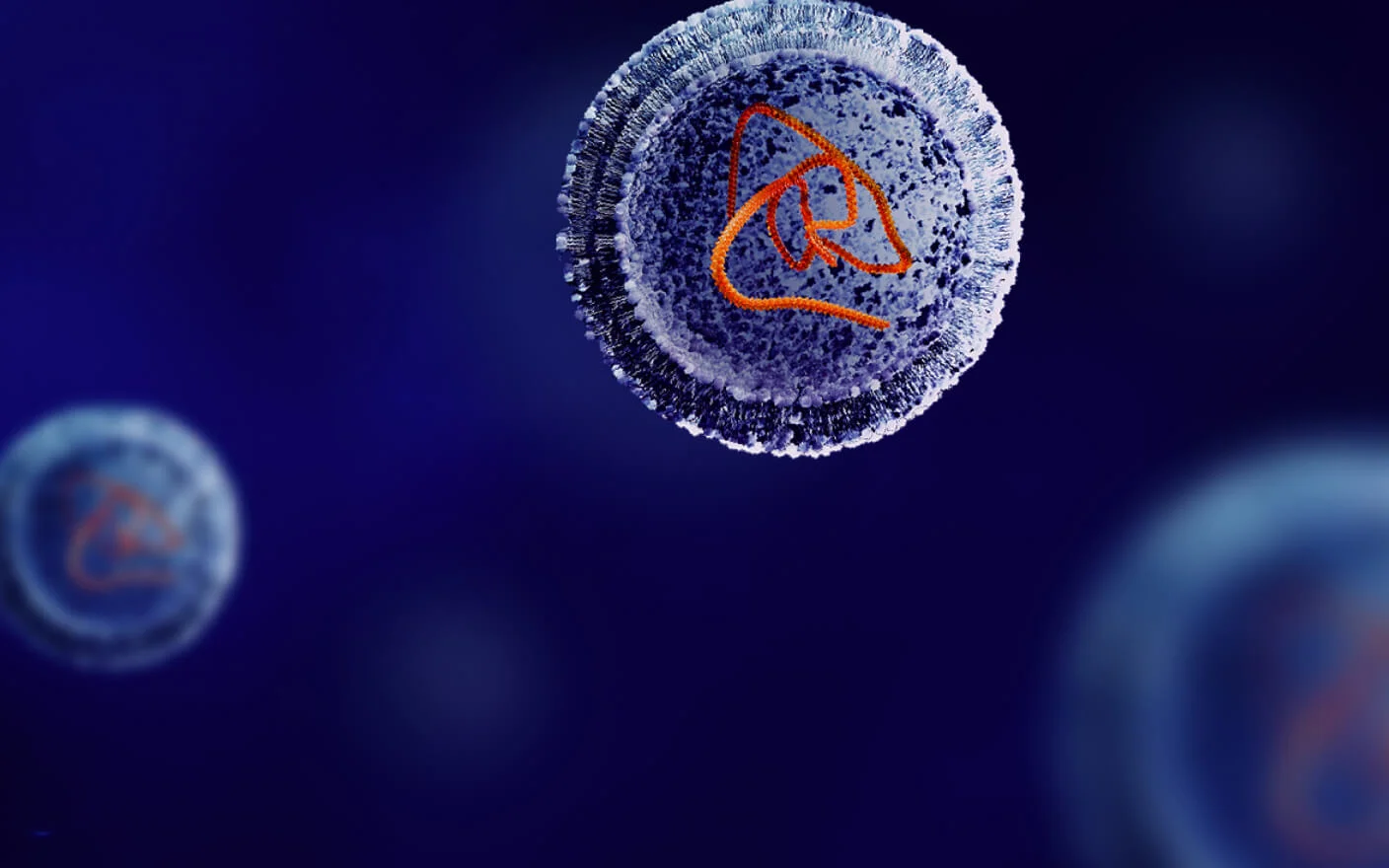
In siRNAs, the strand that is complementary to the target gene mRNA is the antisense strand (AS), while the other strand is the sense strand (SS). As shown in Figure 1, siRNAs enter cells through endocytosis followed by binding with protein factors, such as Argonaute (Ago), forming RNA-induced silencing complexes (RISCs). The AS is retained in the RISC complex, while the SS is cleaved and degraded. siRNAs recognize target gene mRNA through base pairing. Subsequently, Ago uses its endonuclease activity to cleave the target gene RNA, and the resulting RNA fragments are easily degraded by 5′ or 3′ exonucleases and thus silence the target gene at the post-tranional level. As a type of drug with a novel mechanism of action, siRNAs have broad prospects in the treatment of rare diseases, cancer, metabolic disorders, and other diseases. With the approval of the first siRNA drug in 2018, the development of siRNA drugs is constantly on the rise. As of 2023, there are more than 200 siRNA drugs in development worldwide. Non-clinical early in vivo PK studies of siRNAs serve as a vital part of the drug development process, which is used to investigate the absorption and distribution of siRNAs in organisms and establish pharmacodynamics/pharmacokinetics (PD/PK) models. It includes evaluations of siRNA plasma PK, tissue PK, RISC PK, the degradation efficiency of target gene mRNA, and the inhibition efficiency of the target protein. The bioanalysis of siRNAs contains analysis of siRNAs, RISCs, gene expression, and protein levels; therefore, multiple analysis platforms are needed to support the quantitative detection of different analytes.

Figure 1. siRNA-mediated gene silencing mechanism 1
siRNA concentration in plasma/tissue
Most siRNAs are administered via subcutaneous (SC) or intravenous (IV) injection, with a short plasma half-life and reaching maximum concentration within 0.25-2 hours, followed by rapid clearance 2. In early non-clinical in vivo studies, siRNA-related metabolism studies are often lacking. Therefore, to avoid the interference of metabolites on the detection of siRNA concentrations, plasma siRNA concentrations can be quantitatively determined using liquid chromatography–triple quadrupole mass spectrometry (LC-MS/MS). Under denaturing conditions, the double-stranded structure of siRNAs denatures into a sense and antisense strand, which can be detected simultaneously by mass spectrometry with the detection sensitivity of 5–10 ng/mL in plasma. For siRNA coupled with different delivery systems, suitable bioanalytical methods should be selected based on their structures, such as hybrid-based liquid-phase fluorescence detection (LC-FL), ligand binding assay (LBA), and quantitative polymerase chain reaction (qPCR) to meet quantitative requirements.
Tissue distribution of siRNAs
siRNAs are mainly distributed in a limited number of tissues, including the liver, kidney, renal cortex, and lymph nodes, with a high exposure and a long half-life in the liver 2. There is usually a lag of approximately 7–14 days between the plasma peak concentration and the maximum effect of the siRNA drug, and the pharmacokinetics in the plasm and the pharmacodynamics in target tissues do not coincide 3. Therefore, the study of the tissue distribution and tissue PK of siRNA is critical for non-clinical early in vivo PK studies. The concentration of siRNAs in a collected tissue can be quantified using a similar LC-MS/MS method to the analysis method used for plasma samples. Different homogenization schemes need to be used for different types of tissues in tissue homogenates. In addition, imaging methods can be used to study the tissue distribution of labeled siRNAs, such as fluorescence imaging of siRNAs labeled with the fluorophore, Cy5 or Cy7. Imaging methods avoid bleeding and cross-contamination during organ harvesting and preserve the structural information of the tissue, but have limited spatial resolution and lower detection sensitivity than LC-MS/MS methods.
Intracellular RISC concentration
After entering the body, siRNA first needs to bind with intracellular Ago and other protein factors to form RISC. The AS of the double-stranded siRNA is retained, and the SS is degraded rapidly. Subsequently, the AS in the RISC binds with the target mRNA to induce mRNA degradation and thus inhibit protein synthesis. This process may take several hours or even days to reach its peak, resulting in a lag in drug efficacy. Therefore, the concentration of the RISC-siRNA complex is closely related to the drug efficacy of the siRNA. Due to the extremely low content of the RISC-siRNA complex, which may be below 1 ng/g, a more sensitive analysis method is needed for quantitative detection. The detection of RISCs can be achieved by RNA immunoprecipitation coupled with stem-loop reverse tranion-quantitative polymerase chain reaction (RT-qPCR). This method is a derivative of the stem-loop RT-qPCR method to specifically detect siRNAs in the RISC. The method is mainly divided into three steps: firstly, magnetic beads bound with protein A/G are conjugated to an anti-Ago2 antibody followed by specifically capturing the complex of Ago2 and RNA complex. After washing and elution, the siRNA bound to Ago2 is obtained, and it is detected by stem-loop RT-qPCR 4. The sensitivity of this method can reach up to 0.25–1 ng/g.
Target gene mRNA degradation efficiency
The mechanism of action of siRNAs is to induce the degradation of target gene mRNA. Therefore, the level of mRNA degradation directly demonstrates the silencing efficiency of siRNA. The relative expression level of target gene mRNAs was examined by RT-qPCR method with the stable expression of housekeeping genes as reference, in order to detect the degradation efficiency of target mRNA. The study process consists of three steps. First, total RNA is extracted from the tissue and complementary DNA is synthesized under the action of reverse tranase, which is followed by PCR amplification and detection by adding specific primers and detection reagents. There are two common detection methods, including the SYBR Green dye method and the Taqman probe method. SYBR Green is a nucleic-acid-binding dye that has weak fluorescence in the free state. When it is embedded in double-stranded molecules, the fluorescence is greatly enhanced, and this characteristic can be used for quantitative detection. This method has good universality, high sensitivity, economy, and convenience, but the specificity is relatively poor, which may result in non-specific amplification and affect the accuracy of quantification. The principle of the Taqman probe method is that during primer extension, the probe is hydrolyzed, releasing fluorescent groups and thus generating fluorescence for detection. Since the probe and the target sequence are specifically bound, this method has strong specificity and good repeatability, and multiple qPCR analysis can be carried out in a single reaction, which reduces the sampling error. The disadvantage of this method is that the probe needs to be specifically designed for each target gene, resulting in poor universality and higher cost.
Inhibition efficiency of target proteins
siRNAs induce the degradation of target gene mRNA, thus affecting the expression of the target protein. However, protein levels are affected by factors such as protein expression, stability, half-life, and other regulatory factors, and therefore, the degree of the decrease in the target protein level may not necessarily be proportional to the decrease in the mRNA level. Target protein levels can be detected by enzyme-linked immunosorbent assay (ELISA), Western blotting, or LC-MS methods. When using ELISA to detect target proteins, the availability of key reagents needs to be considered. If these reagents are difficult to obtain, Western blotting or LC-MS methods can be used for detection.
2. Case study: integrated bioanalysis of GalNAc-conjugated siRNAs
siRNA-X is an siRNA conjugated with N-acetylgalactosamine (GalNAc) with an asymmetrical RNA/dTdT overhang at the 3′ end of the AS (Figure 2). It targets the C5 complement pathway and treats complement-mediated diseases by inhibiting the hepatic production of C5 complement proteins.

Figure 2. siRNA-X structure
We carried out a PK study in mice according to the conventional early in vivo PK protocol of siRNAs. Plasma, serum, and liver samples were collected and analyzed at multiple time points after a single administration by subcutaneous injection. The specific study design is shown in Figure 3: Three dose groups of low, medium, and high were set, and the mice were tranquilized to collect biological samples at 1 and 6 h and on days 1, 2, 3, 7, 14, and 21 after a single dose.
 Figure 3. siRNA-X mouse PK study design and bioanalytical solutions
LC-MS/MS, liquid chromatography-triple quadrupole mass spectrometry; ELISA, enzyme-linked immunosorbent assay; qPCR, quantitative polymerase chain reaction; siRNA, small interfering RNAs; RISC, RNA-induced silencing complex
In this study, three different bioanalytical platforms and four bioanalytical methods were used to analyze the samples:
1. The plasma and liver concentrations of the siRNA were quantified using LC-MS/MS (Figure 4).
2. Levels of C5 complement protein in serum were detected using ELISA (Figure 5).
3. The RISC concentration in the liver was determined using RNA immunoprecipitation combined with stem-loop RT-qPCR.
4. Target gene expression levels were determined in the liver using stem-loop RT-qPCR.

Figure 4. Determination of the siRNA-X concentration in the livers of mice in different dosage groups by LC-MS/MS

Figure 5. Determination of C5 complement protein levels in serum using enzyme-linked immunosorbent assay (ELISA)
By detecting the RISC concentration in liver tissue and comparing it with the inhibition rate of the target protein or the degradation rate of the target mRNA (Figures 6 and 7), it can be seen that the RISC concentration, rather than the drug concentration in free plasma, is more closely related to the gene silencing effect in target.

Figure 6. RISC concentration in mouse liver vs. C5 complement protein inhibition rate

Figure 7. RISC concentration in mouse liver vs. target gene mRNA degradation efficiency
Conclusion
The Non-GLP Bioanalytical Team of WuXi AppTec possesses comprehensive capabilities in oligonucleotide drug bioanalysis. We have developed and established the five major bioanalytical platforms: LC-MS/MS, liquid chromatography–high-resolution mass spectrometry (LC-HRMS), hybrid-based liquid chromatography–fluorescence detection (LC-FL), ligand binding assays (LBA), and qPCR. These five bioanalytical platforms support oligonucleotide bioanalysis from early drug screening to IND filing, providing diversified bioanalytical solutions. With our extensive experience in oligonucleotide bioanalytical method development, we can deliver high-quality in vitro and in vivo data delivery to accelerate the drug development process. Guided by the principles of extensive technical expertise and exceptional analysis, we focus on building new analytical capabilities. We continue to build a leading non-GLP bioanalytical service with cross-regional collaboration, scale-up, full process integration, and one-stop service advantages.

Figure 8. WuXi AppTec metabolism and PK bioanalytical platforms for oligonucleotides
Click here to learn more about the strategies for oligonucleotides, or talk to a WuXi AppTec expert today to get the support you need to achieve your drug development goals.
Authors: Nan Zhao, Jiaming Jiang, Zhiyu Li, Lili Xing
Committed to accelerating drug discovery and development, we offer a full range of discovery screening, preclinical development, clinical drug metabolism, and pharmacokinetic (DMPK) platforms and services. With research facilities in the United States (New Jersey) and China (Shanghai, Suzhou, Nanjing, and Nantong), 1,000+ scientists, and over fifteen years of experience in Investigational New Drug (IND) application, our DMPK team at WuXi AppTec are serving 1,500+ global clients, and have successfully supported 1,200+ IND applications.
Reference
1 Halib, N.; Pavan, N.; Trombetta, C.; Dapas, B.; Farra, R.; Scaggiante, B.; Grassi, M.; Grassi, G. An Overview of siRNA Delivery Strategies for Urological Cancers. Pharmaceutics 2022, 14, 718.
2 Lade, J.M., Thayer, M., Doherty, D., Basiri, B., Xie, F. and Rock, B.M.(2018), Pharmacokinetics, Metabolism, and Biodistribution of a Liver-Targeted siRNA Therapeutic in Preclinical Species. The FASEB Journal, 32: 833.5-833.5.
3 Jeon, J.Y., Ayyar, V.S. & Mitra, A. Pharmacokinetic and Pharmacodynamic Modeling of siRNA Therapeutics – a Minireview. Pharm Res 39, 1749–1759(2022).
4 Nair JK, H Attarwala , A Sehgal , Q Wang , K Aluri , X Zhang , M Gao , J Liu , R Indrakanti , et al.(2017). Impact of enhanced metabolic stability on pharmacokinetics and pharmacodynamics of GalNAc-siRNA conjugates. Nucleic Acids Res 45:10969–10977.


Related Resources


Related Services and Platforms


-
 DMPK BioanalysisLearn More
-
Novel Drug Modalities DMPK Enabling PlatformsLearn More
-
Novel Drug Modalities BioanalysisLearn More
-
Small Molecules BioanalysisLearn More
-
Bioanalytical Instrument PlatformLearn More
-
PROTAC DMPK ServicesLearn More
-
ADC DMPK ServicesLearn More
-
Oligo DMPK ServicesLearn More
-
PDC DMPK ServicesLearn More
-
Peptide DMPK ServicesLearn More
-
mRNA DMPK ServicesLearn More
-
Covalent Drugs DMPK ServicesLearn More


Stay Connected
Keep up with the latest news and insights.

