

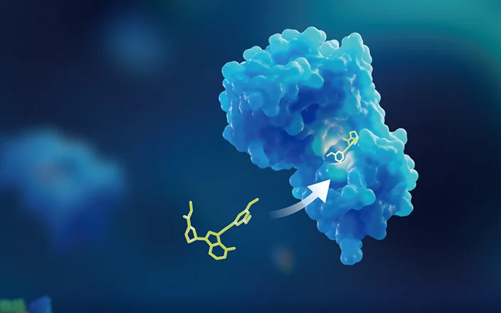
Application of Radiolabeling Techniques in ADC PK Studies
-

Articles
-

Jul 26, 2023
Although some of the payloads of antibody-drug conjugate (ADC) were approved drugs, attention should still be paid to the formation of payload-related catabolites arising from different release mechanisms. It is necessary to study plasma protein binding rate, tissue distribution, metabolism, excretion/mass balance, drug-metabolizing enzymes, and transporter effects, which are commonly conducted for new chemical entries.
Due to low systemic exposure of payloads, severe matrix interference, and the difficulty in predicting the cleavage products of non-cleavable ADCs, studies of tissue distribution, in vivo metabolism, and ADC excretion remains challenging. Literature suggests that ADCs targeting specific tissue lesions need to undergo tissue distribution studies, which can be carried out using radiolabeled payload or by separately radiolabeled antibodies and payloads to obtain more comprehensive ADC tissue distribution characteristics.
This article summarizes the current radiolabeling strategies for ADC drug distribution studies and provides examples of how to screen ADC drugs with the best drug/antibody ratio (DAR) in the drug discovery and development phase and how to apply radiolabeling technology to achieve comprehensive in vivo ADC characterization in their applications.
Radiolabeling Strategies for ADC Drugs
Before performing radioactivity studies, appropriate radioisotopes should be selected for the synthesis of radiolabeled ADC payloads and antibodies separately (Figure 1).
Payloads are usually radiolabeled with low energy radioisotope 14C or 3H, which have specific activities of ~62.4 mCi/mmol (14C) and ~28.6 Ci/mmol (3H), respectively. It is challenging in radioactive detection due to low in vivo concentrations of payload from ADC drugs. It is recommended to increase the radio-labeled drug dose to meet the detection requirements.
Radio-labeled drug dose (µCi/kg) = chemical dose (mg/kg) × specific activity (µCi/mg)
Generally, a higher ADC DAR indicates that more radioisotope has been labeled on a single payload, thus achieving a higher specific activity through synthesis. The 3H-labeled specific activity is significantly higher than that of 14C. However, considering that 3H labeling is prone to hydrogen-tritium exchange in vivo so before and after volatile drying samples need to be tested, increasing the difficulty and cost of each study. Therefore, as long as detection requirements are met, it is recommended to choose 14C to label payloads.
Antibodies are usually radiolabeled with a high-energy radioisotope such as 125I or 89Zr. Because of their high specific activity, the detection limit is no longer a limiting factor. Therefore, factors considered during synthesis include the types of amino acids contained in the antibody (different nuclides are correspondingly labeled with specific amino acids), radioisotope half-life (should not be shorter than the biological half-life of the antibody in vivo), and stability and activity of the post-labeled antibody. 125I is the most common choice for radiolabeling due to its accessibility, simple labeling process, and minimal impact on the activity of the antibody.

Figure 1. Common methods for radiolabeling
*Indicates isotope labeling position
Currently marketed ADC drugs, such as Adcetris and Padcev from Takeda Pharmaceuticals, Kadcyla and Polivy from Roche, Besponsa from Pfizer, Enhertu from AstraZeneca, Zynlonta from ADC Therapeutics, and Tivdak from Seagen/Genmab, all use low-energy radioisotope (14C or 3H) to label payloads for studies of tissue distribution, mass balance, plasma protein binding rate, and metabolite identification. Radioisotopes (125I, 111In, 3H, and 89Zr) were used to label antibodies in Kadcyla, Polivy, Enhertu, Trodelvy, and Tivdak to reveal the difference of ADC tissue distribution in comparison to that observed by using only payload labeled ADC drugs.
Selecting ADC Drugs with Optimal ADC DAR in the Drug Discovery and Development Phase
In the development phase, highly sensitive radiolabeling technology allows clear determination of ADC PK behavior in vivo. In addition, labeling naked antibodies, ADC antibodies, and payloads separately, as well as comparing the difference of in vivo behavior observed by using different labeling locations and conjugation methods could contribute to screening out ADCs that meet the expectations at the earliest possible stages.
SAR3419 is an ADC drug comprising a CD19 protein antibody (huB4) and the microtubule inhibitor DM4 (Maytansine, shortened to May) (Figure 2). A [125I]-labeled Ab was obtained by labeling the naked antibody with 125I, and [125I] Ab-L-May was obtained by labeling the ADC antibody. Administration of [125I] Ab or [125I] Ab-L-May to mice showed identical tissue distributions (Figure 3). This suggests that the in vivo tissue distribution of the naked antibody was not changed after the huB4 antibody was conjugated to the payload DM4 (May)1 with a DAR value between 3.5 and 4.

Figure 2. Constitutional formula of SAR3419 2
![Histogram of tissue distribution in mice after intravenous injection of [125I] Ab (left) and [125I]Ab-L-May (right)](https://wuxiapptec-dmpkcatalog-prod.oss-cn-shanghai.aliyuncs.com//upload/image/20230719/501223278094548.png)
Figure 3. Histogram of tissue distribution in mice after intravenous injection of [125I] Ab (left) and [125I]Ab-L-May (right)1
If adopted antibodies are identical, do different DAR values affect the in vivo distribution of payloads? In the study design shown below, after synthesis of [3H] payloads, cleavable (M9346A-sulfoSPDB-DM4) and non-cleavable (J2898A-SMCC-DM1) ADCs were prepared with DAR values ranging from low (mean »2, range: 0–4) to very high (mean »10, range: 7–14). PK analyses in mice showed that ADC clearance was slow for DAR means below six and became significantly faster for DAR means above nine. Tissue distribution results indicated that tissue distribution characteristics of ADC with lower DAR were similar to those of naked antibodies, while tissue distribution characteristics of ADC with DAR above nine differed significantly from those of naked antibodies. Payload concentrations declined rapidly in whole blood after dosing, accumulated rapidly in the liver, and were rapidly excreted via the hepatic routes, resulting in limited in vivo exposure and subsequently suboptimal pharmacodynamic performance (Figures 4 and 5).2
![Pharmacokinetic study in CD1 mice after intravenous injection of M9346A-sulfo-SPDB-[3H]DM4 (A) and J2898A-SMCC-[3H]DM1 (B)](https://wuxiapptec-dmpkcatalog-prod.oss-cn-shanghai.aliyuncs.com//upload/image/20230719/468471416512905.png)
Figure 4. Pharmacokinetic study in CD1 mice after intravenous injection of M9346A-sulfo-SPDB-[3H]DM4 (A) and J2898A-SMCC-[3H]DM1 (B)2
![Tissue distribution study of CD1 mice after intravenous injection of 10 mg/kg M9346A-sulfo-SPDB-[3H]DM4 (A) and J2898A-SMCC-[3H]DM1 (B)](https://wuxiapptec-dmpkcatalog-prod.oss-cn-shanghai.aliyuncs.com//upload/image/20230719/492488900223176.png)
Figure 5. Tissue distribution study of CD1 mice after intravenous injection of 10 mg/kg M9346A-sulfo-SPDB-[3H]DM4 (A) and J2898A-SMCC-[3H]DM1 (B)2
In the Application Phase, Using Radiolabeling Techniques to Profile the In Vivo Characteristics of ADC Drug
Tissue distribution studies are necessary for ADCs targeting specific tissues lesion and they should be conducted using pharmacodynamic animal models. It is important to highlight that quantitative whole-body autoradiography (QWBA) can provide average concentrations within entire tissues and can distinguish concentration differences among regions within the tissue and then quantify tissues by region. Compared with conventional harvesting methods, QWBA offers incomparable advantages in assessing dose-response relationships between the ADC distribution depth in tumor tissues and drug efficacy.
Trastuzumab deruxtecan (code name, DS-8201a or T-DXd; US brand name, Enhertu), launched in 2019, is a combination of a HER2-targeting antibody and Dxd, a DNA topoisomerase I inhibitor, for treating adult patients with HER2-positive unresectable or metastatic breast cancer. Its PK studies are documented in published literature, as described below and illustrated in Figure 6.

Figure 6. Constitutional formula and radiolabeling position in DS-8201a
To distinguish different radiolabeling positions, the ADC is represented as Ab-L-Dxd (Ab: antibody; L: linker; Dxd: small molecule). The antibodies of 3H-labeled ADCs are denoted as [3H] Ab-L-Dxd and those of 14C-labeled payload are represented as Ab-L-[14C] Dxd. Additionally, 14C labeled payload only is indicated as [14C] Dxd.
Two groups of cynomolgus monkeys were administered [3H] Ab-L-Dxd or Ab-L-[14C] Dxd, respectively, and the differences in tissue distribution between them were assessed using QWBA. The results showed that the distribution characteristics of the two ADCs with different radiolabeling locations were similar in whole blood and important tissues and organs. The only difference was that Ab-L-[14C] Dxd exhibited a high distribution in the large intestine, while [3H] Ab-L-Dxd displayed a low distribution in the intestinal tract. The main excretion pathway of Ab-L-[14C] Dxd was feces (67.3%), and the secondary excretion pathway was urine (18.7%). No metabolites other than [14C] Dxd were detected in feces and urine. In addition, bile duct cannulation studies in rats showed that 71.5% of the unconjugated payload [14C] Dxd was excreted via the bile (Figures 7–9).
The study results from the two animal models described above provide a comprehensive understanding of the in vivo pharmacokinetic process of Ab-L-Dxd: after Ab-L-Dxd is injected into the body, following the antibody to distribute throughout the body, with the highest concentration in whole blood. It does not accumulate in normal tissues, which considerably reduces its safety risk. After the release of Dxd in the body, Dxd is basically not metabolized and most of it flows into the intestinal lumen via the bile and is eventually excreted from the feces.3
![Representative quantitative whole-body autoradiography images at 24 h (a) (c) and 336 h (b) (d) after intravenous injection of [3H] Ab-L-Dxd (a) (b) and Ab-L-[14C] Dxd (c) (d) in cynomolgus monkeys](https://wuxiapptec-dmpkcatalog-prod.oss-cn-shanghai.aliyuncs.com//upload/image/20230719/560789247031370.png)
Figure 7. Representative quantitative whole-body autoradiography images at 24 h (a) (c) and 336 h (b) (d) after intravenous injection of [3H] Ab-L-Dxd (a) (b) and Ab-L-[14C] Dxd (c) (d) in cynomolgus monkeys.3
![The 0–336 h cumulative excretion percentage after intravenous injection of Ab-L-[14C]Dxd in cynomolgus monkeys, and the 0–48 h cumulative excretion percentage after intravenous injection of [14C]Dx](https://wuxiapptec-dmpkcatalog-prod.oss-cn-shanghai.aliyuncs.com/Public/Uploads/ueditor/upload/image/20241025/1729825874617176.jpg "1729825874617176.jpg")
Figure 8. The 0–336 h cumulative excretion percentage after intravenous injection of Ab-L-[14C]Dxd in cynomolgus monkeys, and the 0–48 h cumulative excretion percentage after intravenous injection of [14C]Dxd in rats with bile duct intubation3.
![Radioactive metabolite profiles in feces and urine after intravenous injection of Ab-L-[14C] Dxd in cynomolgus monkeys](https://wuxiapptec-dmpkcatalog-prod.oss-cn-shanghai.aliyuncs.com/Public/Uploads/ueditor/upload/image/20241025/1729825942536930.jpg "1729825942536930.jpg")
Figure 9. Radioactive metabolite profiles in feces and urine after intravenous injection of Ab-L-[14C] Dxd in cynomolgus monkeys
Comprehensive analysis of filing information and relevant literature on the pharmacokinetics of approved ADCs, combined with experimental experience, it is recommended that in vitro study as the first step of ADC DMPK studies, including the metabolite identification of payload related to ADC release in liver S9 or tumor cells, and studies of plasma/serum stability of ADCs. Based on the results of in vitro assay, it is possible to make a preliminary determination of whether new unconjugated payloads are released from ADC.
In animal tests, plasma concentration and tissue distribution results are essential for ADC efficacy and safety assessments, and concentrations of ADC, total antibody, and unconjugated payloads should be assessed. It is recommended to adopt radiolabeling techniques to track the tissue distribution in vivo, and to examine whether the tissue distribution characteristics of radiolabeled payloads and antibodies are similar. If a significant difference is observed in a particular tissue, the specific reason can be inferred by the in vivo behavior of the unconjugated payload. Subsequently, based on metabolite identification results from radioactive plasma, urine, and feces, it can be further confirmed whether an ADC releases payload-related catabolites in vivo. The supplementary plasma protein binding ratio, drug-metabolizing enzymes, and transporter effects studies of the payload-related catabolites should be conducted following guideline requirements.
Conclusion
By incorporating regulatory approval request data, literature studies, and extensive experience, radiolabeling techniques allow for more precise tissue distribution assessments of ADC drugs. In different drug development phases, radiolabeling techniques can elucidate the fundamental characteristics of a drug and aid in the selection of the most promising candidates.
Click here to learn more about the strategies for ADC, or talk to a WuXi AppTec expert today to get the support you need to achieve your drug development goals.
Authors: Xue Yu, Huan Li, Lingling Zhang
Committed to accelerating drug discovery and development, we offer a full range of discovery screening, preclinical development, clinical drug metabolism, and pharmacokinetic (DMPK) platforms and services. With research facilities in the United States (New Jersey) and China (Shanghai, Suzhou, Nanjing, and Nantong), 1,000+ scientists, and over fifteen years of experience in Investigational New Drug (IND) application, our DMPK team at WuXi AppTec are serving 1,500+ global clients, and have successfully supported 1,200+ IND applications.
Reference
[1] Hans K. Erickson1,2 and John M. Lambert. ADME of Antibody–Maytansinoid Conjugates. The AAPS Journal, Vol. 14, No. 4, December 2012 (# 2012)
[2] Xiuxia Sun, Jose F. Ponte, Nicholas Yoder, et al. Effects of Drug-Antibody Ratio (DAR) on Pharmacokinetics, Biodistribution, Efficacy and Tolerability of Antibody-Maytansinoid Conjugates.
Bioconjug Chem. 2017 May 17;28(5):1371-1381.
[3] Yoko Nagai, Masataka Oitate, Hideyuki Shiozawa, et al. Comprehensive preclinical pharmacokinetic evaluations of trastuzumab deruxtecan (DS-8201a), a HER2-targeting antibody-drug conjugate, in cynomolgus monkeys, Xenobiotica, 49:9, 1086-1096,


Related Services and Platforms


-
 Radiolabeled In Vivo ADME StudyLearn More
-
Novel Drug Modalities DMPK Enabling PlatformsLearn More
-
Radiolabeled MetID (Metabolite Profiling and Identification)Learn More
-
Radiolabeled Non-Clinical In Vivo ADME StudyLearn More
-
Quantitative Whole-body Autoradiography (QWBA)Learn More
-
Human Radiolabeled Mass Balance StudyLearn More
-
Radiolabeled Compound SynthesisLearn More
-
PROTAC DMPK ServicesLearn More
-
ADC DMPK ServicesLearn More
-
Antibody-Oligonucleotide Conjugate (AOC) DMPK ServicesLearn More
-
Oligo DMPK ServicesLearn More
-
PDC DMPK ServicesLearn More
-
Peptide DMPK ServicesLearn More
-
mRNA DMPK ServicesLearn More
-
Covalent Drugs DMPK ServicesLearn More


Stay Connected
Keep up with the latest news and insights.

